Art of Science 8.0
April 10, 2018 - April 10, 2018
CURATED BY
Noah Dibert
WHEN
November 30 - November 30
WHERE

View Gallery
1
/
10
1 / x

Order and Light
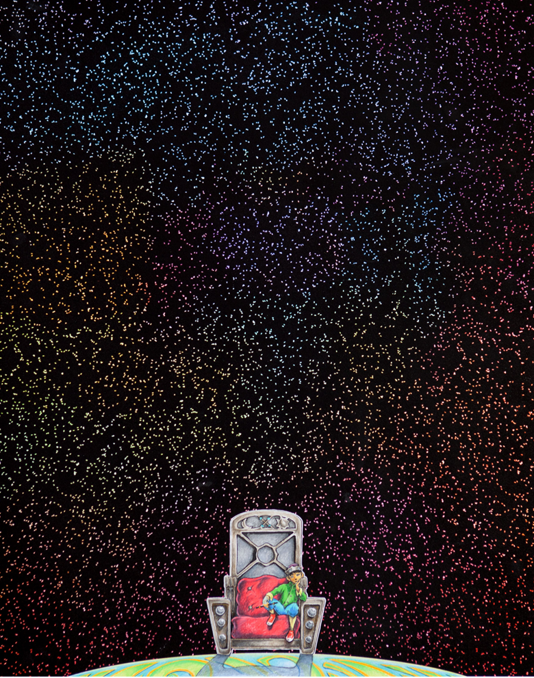
Much like some of the brighter stars in our night sky, a closer look might reveal that the points of light scattered across this image are actually pairs of bright spots, not singular ones. They are created by attaching two dye molecules to specially designed DNA fragments that fold origami-like into a standardized structure. Being able to image the two bright spots distinctly on this “ruler” tells microscopists the resolution of their instrument.
Whorl of the World
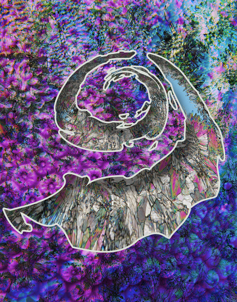
Scales? Feathers? Iridescent fabric? These delicate patterns are skeletons of corals, composed of the mineral aragonite, which typically has needle- and fan-shaped crystals. Through an innovation in microscopy that looks at light in two different ways as it illuminates the same sample, these structures are revealed in colorful images like this one. The study of corals provides scientists with a sensitive indicator of past and present climate around the planet.
Magia Naturalis
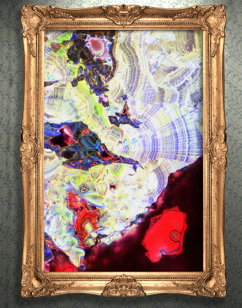
This stone, shown in cross-section, was formed with the aid of microbes in a warm, dark, salty hot spring—the inside of a human kidney. The multiple colors and sharp lines mark where the stone dissolved and recrystallized, fractured and reformed over time, trapping organic matter inside as it came back together. By comparing stone formation in geological and medical settings, researchers are gaining new insights into kidney stone treatment and prevention.
Bee Larvae
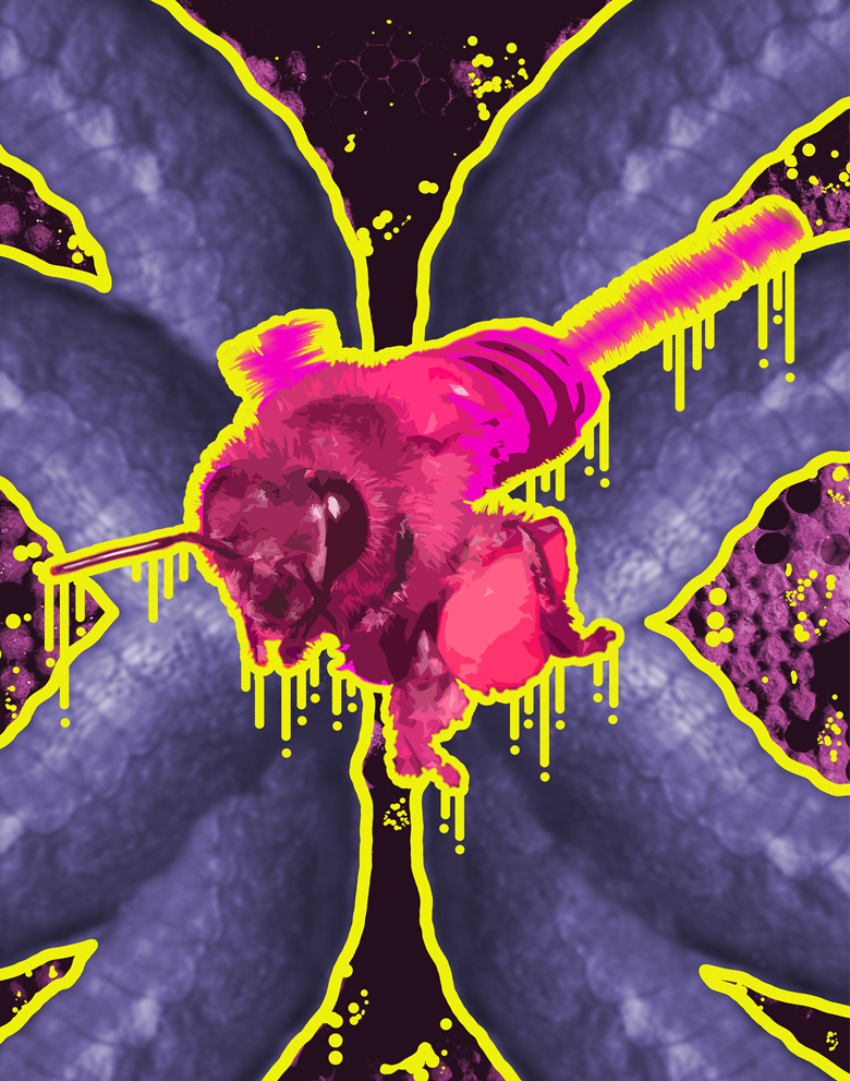
Honey bee pollination helps to ensure the availability of many agricultural crops. Food security is threatened by a worldwide decline in honey bee populations due to four main factors: parasites, pathogens, pesticides and poor nutrition. Researchers are learning how to use advanced robotics to rear honey bee larvae in the laboratory, like those seen in the background of this image, to produce new colonies and study how these four factors interact to affect bee health.
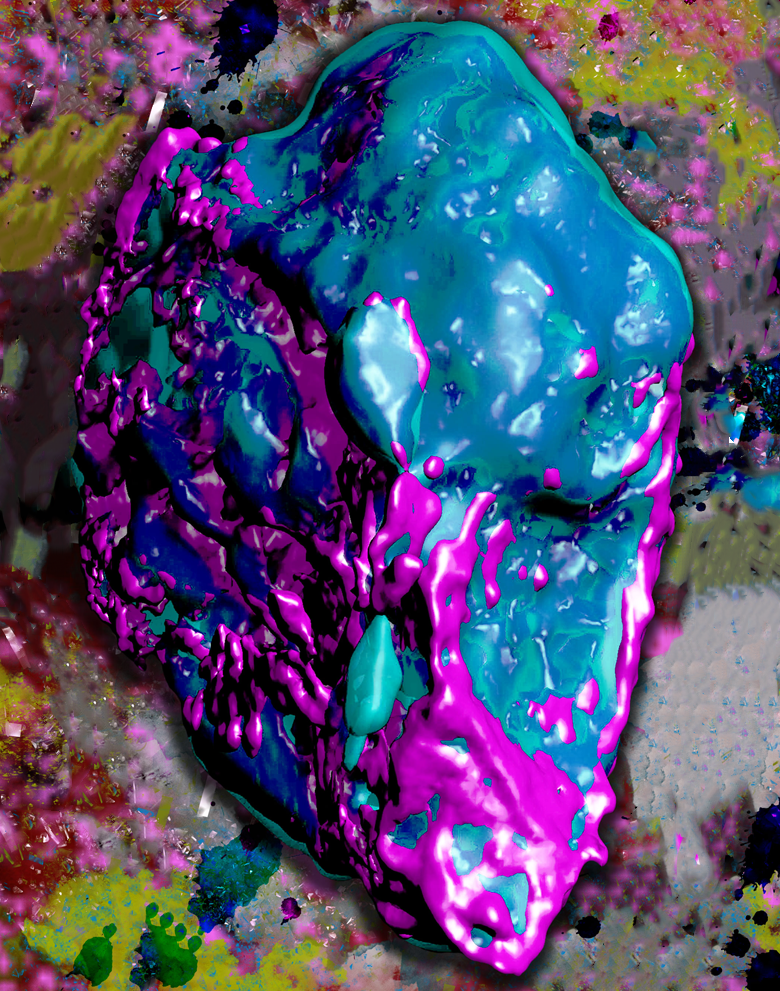
Time's Treasury
This image shows a piece of jasperoid, a rare type of rock made up of quartz and iron oxide crystallites. The varying colors seen here represent the presence of different elements within the rock; images like this one help researchers track the decay of different components and estimate a sample’s age. That information can help inform the search for useful minerals, the study of ancient environments, and even the understanding of geological events on other planets.
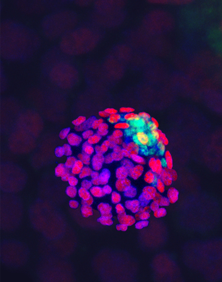
Genesis
This amorphous clump of cells is actually a mouse embryo in one of the earliest stages of development. The rainbow of colors mark different cell components and types within the embryo; by visually identifying them, scientists can track the effects of exposure to MEHP, a chemical used in plastics manufacture that can interfere with hormonal signaling and might impact healthy development in utero.
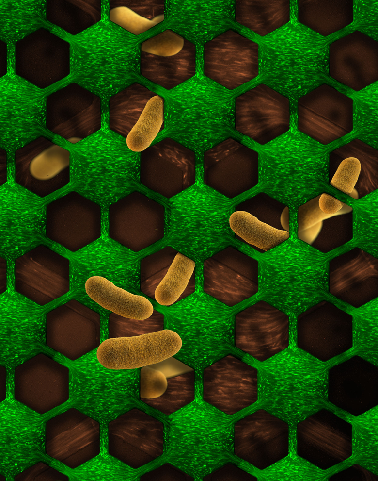
Tipping Point
A common bacterial species in both the human gut and biological laboratories is Escherichia coli, whose individual rod-like shapes can be seen here in extreme close-up and as tiny points of green light. The cells are flowing through a microfluidic chamber that allows researchers to observe how they respond to different concentrations of an antibiotic drug. Their dynamic responses reveal how bacteria in nature react when exposed to stresses and can help reveal the processes that lead to antibiotic resistance.
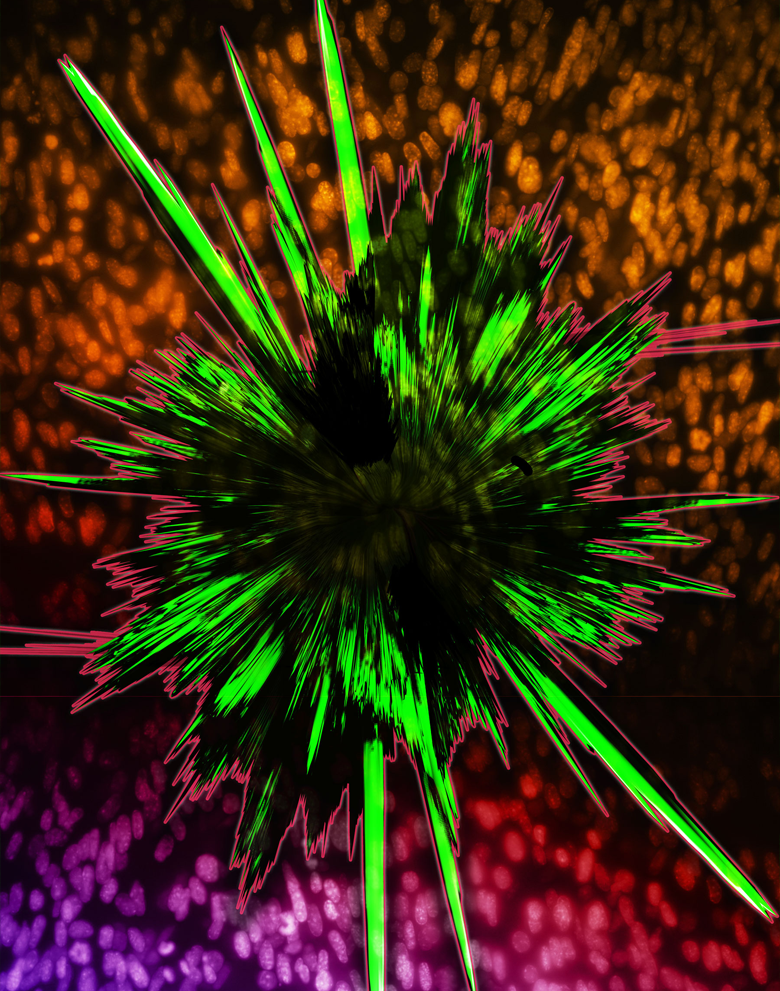
Breaking the Plastic Ceiling
Much like some of the brighter stars in our night sky, a closer look might reveal that the points of light scattered across this image are actually pairs of bright spots, not singular ones. They are created by attaching two dye molecules to specially designed DNA fragments that fold origami-like into a standardized structure. Being able to image the two bright spots distinctly on this “ruler” tells microscopists the resolution of their instrument.
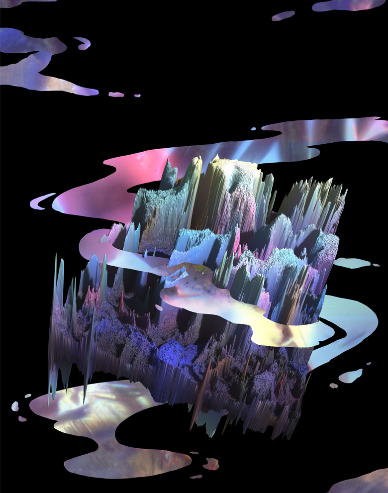
Castle on a Cloud
This mountain ridge in miniature was formed by calcium oxalate crystals found in a thin section of a kidney stone. Most kidney stones are made of calcium oxalate; not all calcium oxalate crystals are made alike. Each type of crystal will incorporate mineral and organic matter at different rates, making kidney stone formation much more complex than scientists originally believed it to be. Studying how these crystals form will help researchers develop new ways to prevent and treat this painful condition.
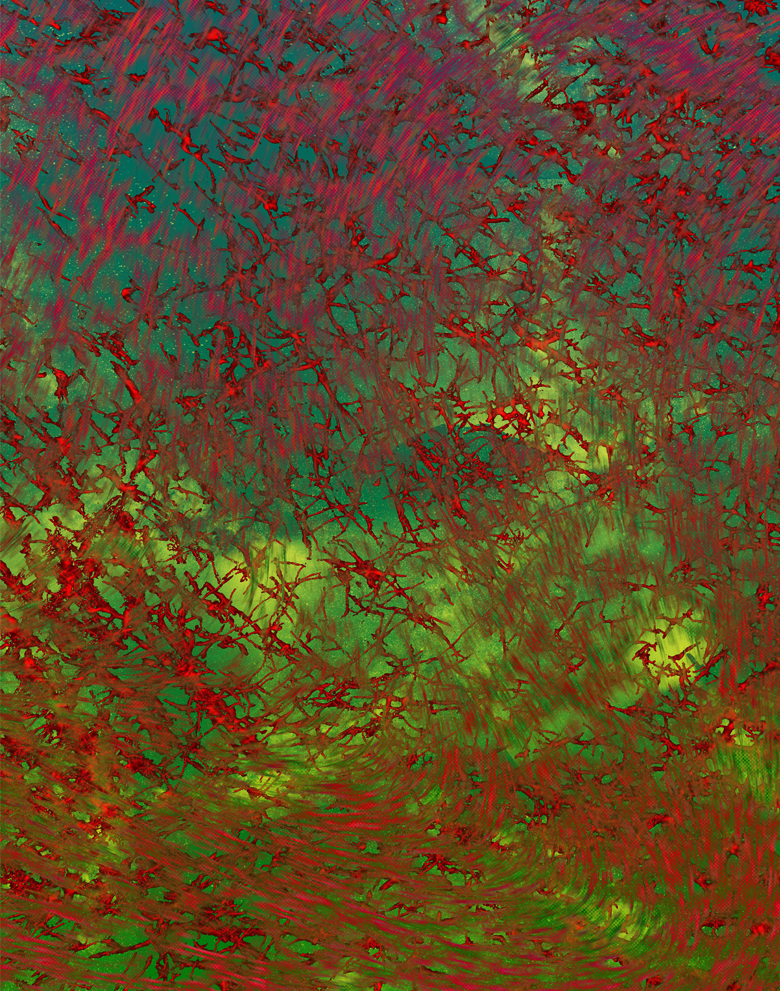
The Bartlet Proxy
Multiple sclerosis is a progressive, disabling illness in which the body’s immune system attacks myelin, an important tissue of the nervous system. Myelin acts like insulation around nerve cells, helping them to conduct the electrochemical signals they use to communicate. Researchers use images like this one, which reveals the web-like structure of myelin in the mouse brain, to study how environmental factors such as viral infection might contribute to the disease process of multiple sclerosis and suggest better treatments.
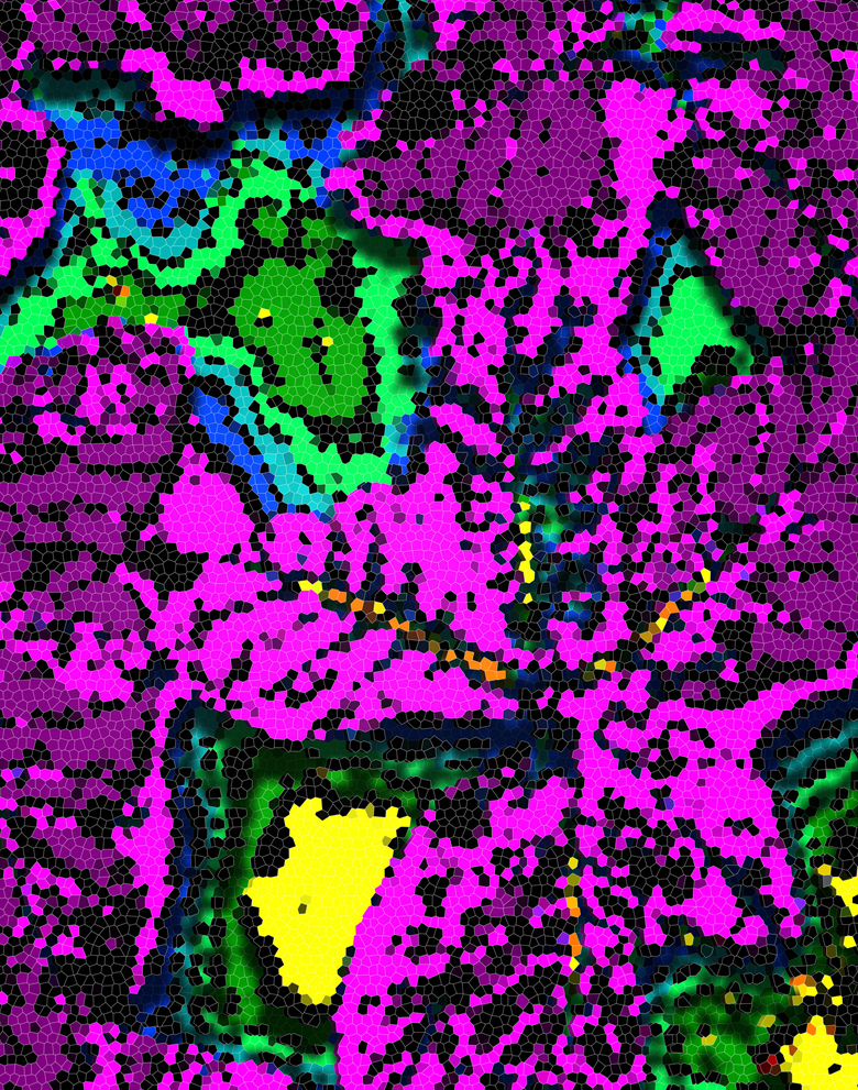
Solar Panels
This patchwork of colors is a thermal image of tobacco plants taken at noon during mid-July. The leaf veins dominate the image foreground, while the warm soil provides the image’s backdrop. These plants have very large leaves, which means the plant has more surface area in between the veins to transpire or “sweat,” keeping the leaves cooler. Researchers are interested in the processes that keep leaves cool and preserve more moisture to inform the development of water-efficient and drought-resistant crops.
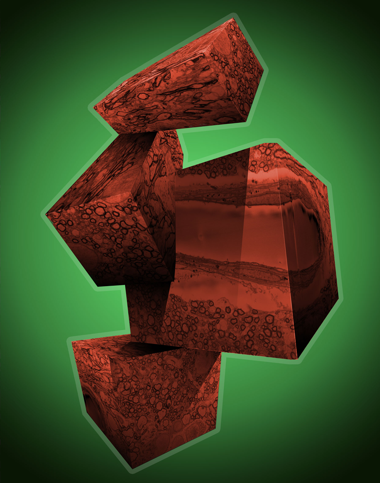
Building Blocks
This image shows the composition of the brain on a cellular level: one can see the half-pipe shape of a blood vessel wall and the smaller cross-sections of neurons and other brain cells. Researchers are investigating the anatomy of the pig brain to better understand how early-life nutrition affects brain development. By studying an animal model with very similar neuroanatomy as the human, researchers hope to improve upon infant formula and its ability to promote health.
Circles of Life
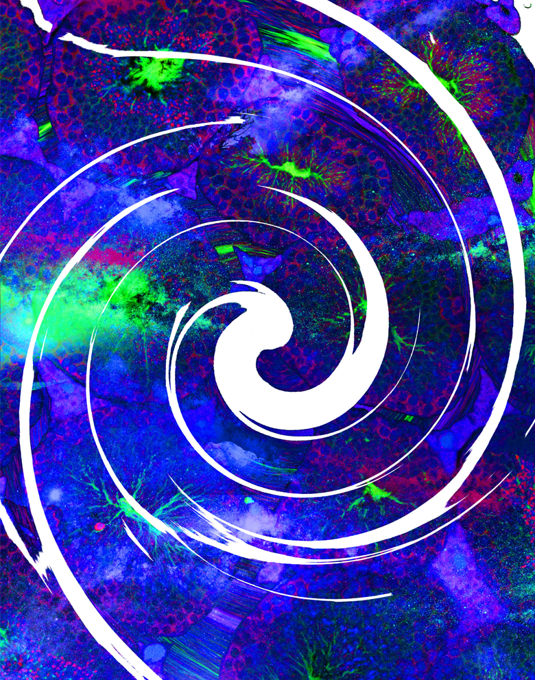
New life could not be created without the process that happens inside these colorfully labeled structures. The small round shapes in this image of mouse tissue are formed by Leydig cells, which produce male steroid hormones within the testes. These are contained by the seminiferous tubules, seen here in cross-section, within which the process of spermatogenesis, the production of individual sperm cells, occurs. Understanding of healthy and disordered fertility is aided by anatomical studies that rely on images like this one.
Cognitive Map
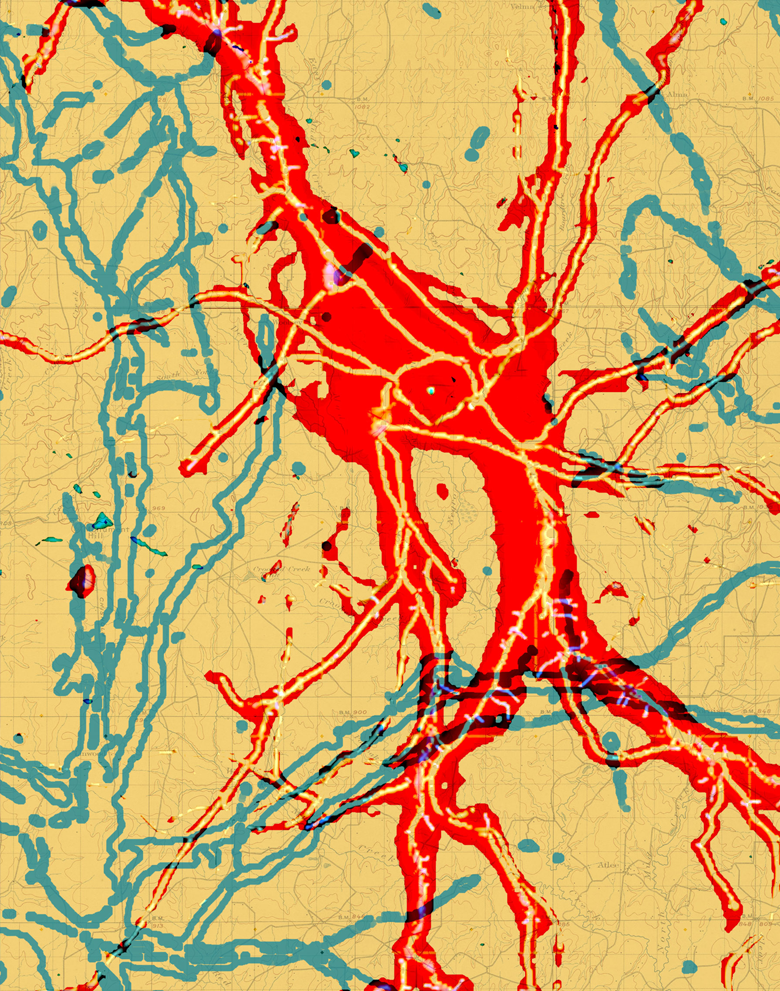
The meandering tracks across this image trace the shape of an astrocyte, a specialized type of brain cell. Astrocytes were once thought to simply support neurons, but have recently become recognized as a more central player in regulating processes in the brain. The structural complexity of astrocytes is a possible factor that affects neuronal function; more complex astrocytes are more likely involved in complex neuronal functions.
The Power of Perspective
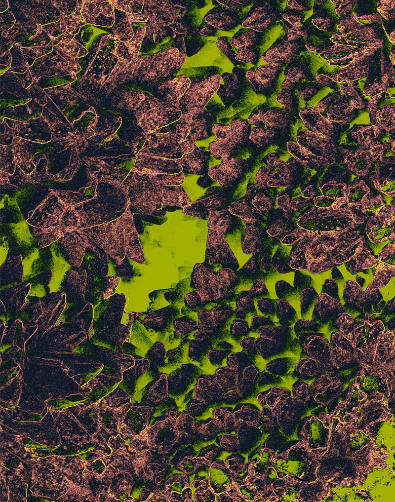
This image might make you think of grains of sand, a boulder-strewn landscape, or a mountain range viewed from above. These shapes are actually found at the opposite end of the scale: they are microscopic calcite crystals in a hot-spring travertine deposit that grew from ancient melting glacier waters flowing through what is now Yellowstone National Park. By studying microbial influences on mineral depositions such as this one, researchers can gain information to improve energy production, human medicine, and even space exploration.
Under Construction
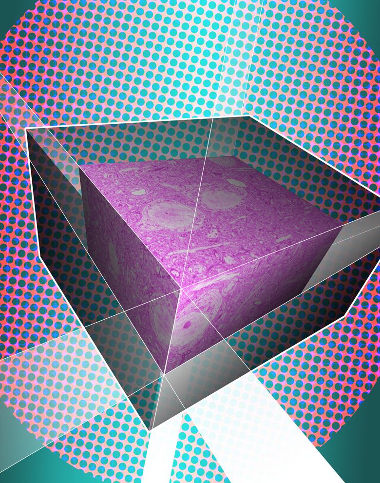
The central focus of this image is a 3D reconstruction of the connections between the two major cell types in the brain, neuron, and glia. As the brain develops, connections between cells are constantly forming, changing, and disappearing as new information and motor skills are learned. Difficulties with motor skill learning are common in children with autism and other developmental disorders; a close investigation of how these connections form and change during motor skill learning can help us better understand these disorders.
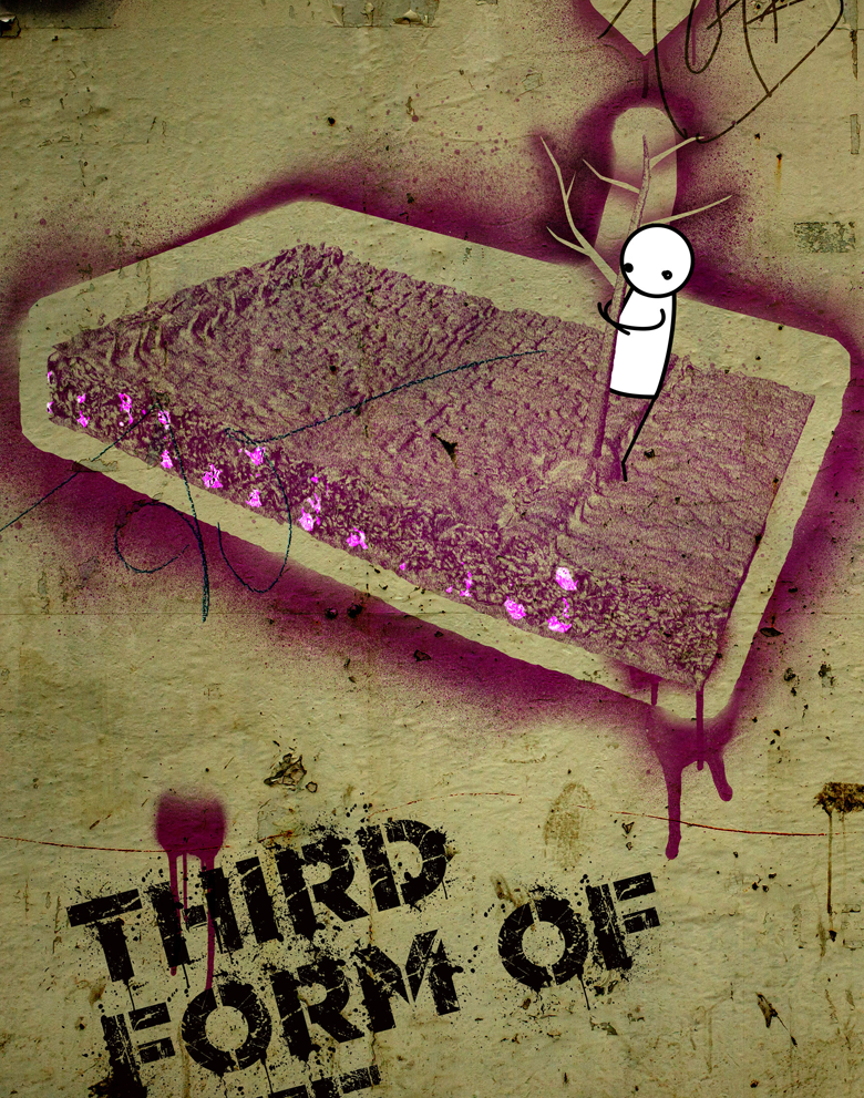
The Smallest Patch of Green
The seemingly subterranean glow seen here actually marks air cavities within the leaf of a maize plant, beneath the pores on the surface of the leaf that allow the plant to take in carbon dioxide and release water vapor. The more area within these cavities, the more the leaves can breathe. Images like this one help plant scientists explore the photosynthetic properties of different varieties of plants.
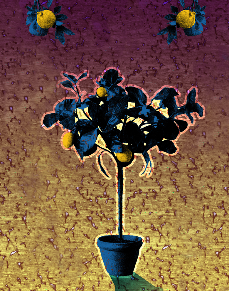
Lemons Aid
Pectin may be familiar as the type of sugar that gives jams and candies their pleasantly gelatinous texture. In the background of this image, you can see what pectin molecules, which are often derived from citrus fruits, look like up close. These molecules link together to form the structure that makes foods gel. These molecules can also be modified to give foods different properties.
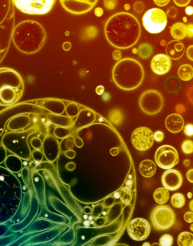
Aerein
Our lungs expand and contract when we breathe. Surfactants, complex mixtures of chemicals produced by our bodies, allow lungs to inflate more easily after exhalation; they prevent the smaller air sacs from collapsing, and the larger ones from being distended during inhalation. With images like this one, which shows little bubbles made with surfactant, researchers are studying the composition of surfactant and how it functions in healthy and diseased lungs.
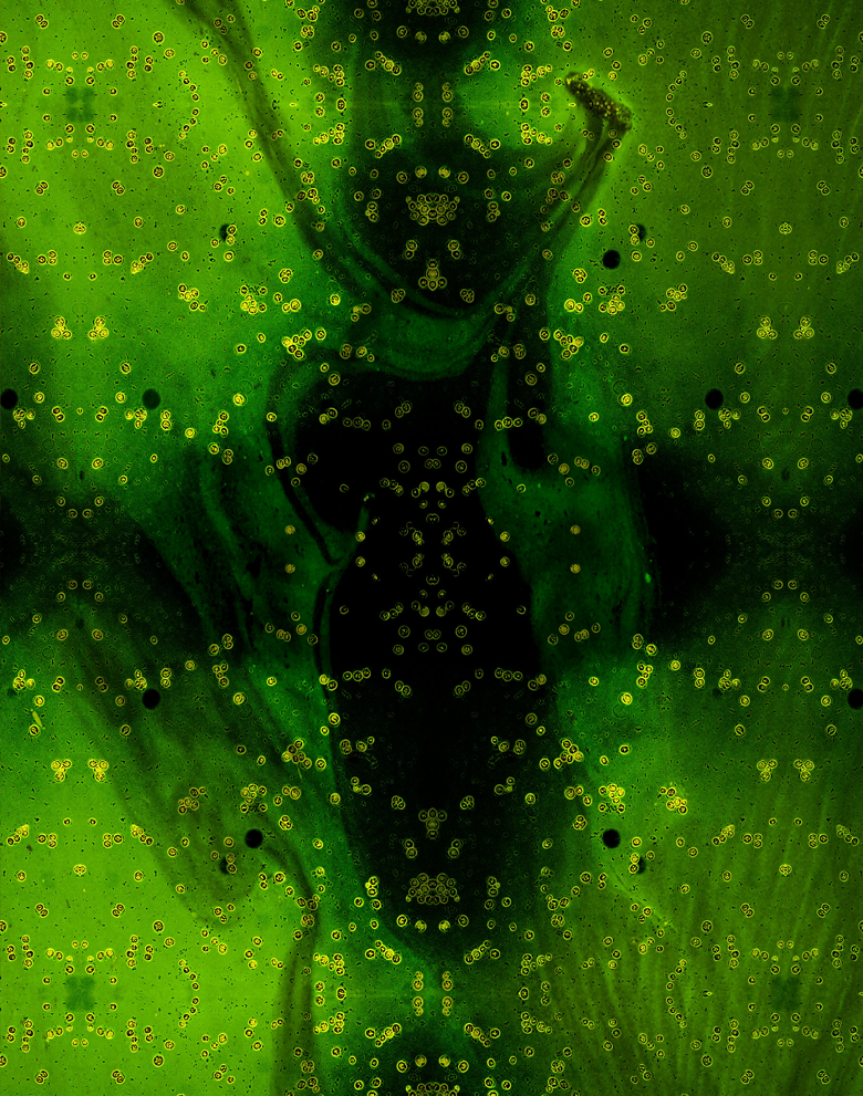
Living in a Shadow
The single-celled green algae seen in this image have a special ability: they can photosynthesize, converting light into food as plants do, but can also eat and grow in the dark if provided with sugar. Researchers are studying how this versatile organism is affected by the chemical nature of the environment using new microfluidic technology. This will help throw light on the impact of human disturbances of ecosystems and how they can be mitigated.

Nanoruler
Much like some of the brighter stars in our night sky, a closer look might reveal that the points of light scattered across this image are actually pairs of bright spots, not singular ones. They are created by attaching two dye molecules to specially designed DNA fragments that fold origami-like into a standardized structure. Being able to image the two bright spots distinctly on this “ruler” tells microscopists the resolution of their instrument.
Order and Light
Scientist Collaborator
Mayandi Sivaguru
IGB Core Facilities
Instrument
Zeiss Elyra S1 Super Resolution Structured Illumination Microscope
Funding Agency
Funded by the University of Illinois
Original Imaging


Image Rights
Images not for public use without permission from the Carl R. Woese Institute for Genomic Biology.
Share
Whorl of the World
Scientist Collaborator
Mayandi Sivaguru, Kyle Fouke, Mike Kingsford and Bruce Fouke
Bruce Fouke Laboratory
Instrument
Nanozoomer Slide Scanner
Funding Agency
Funded by NASA
Original Imaging


Image Rights
Images not for public use without permission from the Carl R. Woese Institute for Genomic Biology.
Share
Magia Naturalis
Scientist Collaborator
Jessica Saw and Mayandi Sivaguru
Bruce Fouke and Derek Wildman Laboratories
Instrument
Zeiss LSM 880 Microscope with Airyscan
Funding Agency
Funded by Mayo Clinic, the Mayo Clinic and University of Illinois Strategic Alliance for Technology-Based Healthcare, and NASA
Original Imaging


Image Rights
Images not for public use without permission from the Carl R. Woese Institute for Genomic Biology.
Share
I Will Work Harder
Scientist Collaborator
Julia Fine, William Streyer, Ran Chao and Nathan Beach
Gene Robinson Laboratory
Instrument
Zeiss Axiozoom V16 Microscope
Funding Agency
Funded by the United States Department of Defense
Original Imaging


Image Rights
Images not for public use without permission from the Carl R. Woese Institute for Genomic Biology.
Share
Time's Treasury
Scientist Collaborator
Willy Guenthner
Willy Guenthner Laboratory
Instrument
Micro-CT; Imaris 3D visualization software
Funding Agency
Funded by the National Science Foundation
Original Imaging


Image Rights
Images not for public use without permission from the Carl R. Woese Institute for Genomic Biology.
Share
Genesis
Scientist Collaborator
Rachel Braz Arcanjo
Romana Nowak Laboratory
Instrument
Zeiss LSM 880 Microscope with Airyscan
Funding Agency
Funded by the National Institutes of Health and Science Without Borders
Original Imaging


Image Rights
Images not for public use without permission from the Carl R. Woese Institute for Genomic Biology.
Share
Tipping Point
Scientist Collaborator
Jinzi Deng
Bruce Fouke Laboratory
Instrument
Zeiss Axiozoom V16 Microscope
Funding Agency
Funded by NASA
Original Imaging


Image Rights
Images not for public use without permission from the Carl R. Woese Institute for Genomic Biology.
Share
Breaking the Plastic Ceiling
Scientist Collaborator
Gelson J. Pagan-Diaz
Rashid Bashir Laboratory
Instrument
Zeiss Lightsheet Z.1 Microscope
Funding Agency
Funded by the National Science Foundation
Original Imaging


Image Rights
Images not for public use without permission from the Carl R. Woese Institute for Genomic Biology.
Share
Castle on a Cloud
Scientist Collaborator
Jessica Saw and Mayandi Sivaguru
Bruce Fouke and Derek Wildman Laboratories
Instrument
Zeiss LSM 880 Microscope with Airyscan
Funding Agency
Funded by Mayo Clinic, the Mayo Clinic and University of Illinois Strategic Alliance for Technology-Based Healthcare, and NASA
Original Imaging


Image Rights
Images not for public use without permission from the Carl R. Woese Institute for Genomic Biology.
Share
The Bartlet Proxy
Scientist Collaborator
Emily Ryder
Andrew Steelman Laboratory
Instrument
Zeiss Sigma VP 3View Serial Block-Face Scanning Electron Microscope; Imaris 3D visualization software
Funding Agency
Funded by the University of Illinois
Original Imaging


Image Rights
Images not for public use without permission from the Carl R. Woese Institute for Genomic Biology.
Share
Solar Panels
Scientist Collaborator
Evan Dracup
Carl Bernacchi Laboratory
Instrument
FLIR T1030sc Infrared Camera
Funding Agency
Funded by the Bill & Melinda Gates Foundation
Original Imaging


Image Rights
Images not for public use without permission from the Carl R. Woese Institute for Genomic Biology.
Share
Building Blocks
Scientist Collaborator
Stephen Fleming and Kingsley Boateng
Ryan Dilger Laboratory
Instrument
Zeiss Sigma VP 3View Serial Block-Face Scanning Electron Microscope
Funding Agency
Funded by Mead Johnson Nutrition
Original Imaging


Image Rights
Images not for public use without permission from the Carl R. Woese Institute for Genomic Biology.
Share
Circles of Life
Scientist Collaborator
Kailiang Adam Li
Romana Nowak Laboratory
Instrument
Zeiss 710 Multiphoton Confocal Microscope
Funding Agency
Funded by the National Institutes of Health
Original Imaging


Image Rights
Images not for public use without permission from the Carl R. Woese Institute for Genomic Biology.
Share
Cognitive Map
Scientist Collaborator
Shah Tauseef Bashir
Martha Gillette Laboratory
Instrument
Zeiss LSM 880 Microscope with Airyscan; Imaris 3D visualization software
Funding Agency
Funded by the University of Illinois
Original Imaging


Image Rights
Images not for public use without permission from the Carl R. Woese Institute for Genomic Biology.
Share
The Power of Perspective
Scientist Collaborator
Bruce Fouke
Bruce Fouke Laboratory
Instrument
Zeiss LSM 880 Microscope with Airyscan; Imaris 3D visualization software
Funding Agency
Funded by NASA
Original Imaging


Image Rights
Images not for public use without permission from the Carl R. Woese Institute for Genomic Biology.
Share
Under Construction
Scientist Collaborator
Kingsley Boateng, Tom Bargar, and Anna Dunaevsky
Anna Dunaevsky Laboratory
Instrument
Zeiss Sigma VP 3View Serial Block-Face Scanning Electron Microscope
Funding Agency
Funded by the National Institutes of Health
Original Imaging


Image Rights
Images not for public use without permission from the Carl R. Woese Institute for Genomic Biology.
Share
The Smallest Patch of Green
Scientist Collaborator
Jiayang Xie
Andrew Leakey Laboratory
Instrument
Micro-CT; Imaris 3D visualization software
Funding Agency
Funded by the National Science Foundation
Lemons Aid
Scientist Collaborator
Wenjun Wang
Hao Feng and Donghong Liu Laboratories
Instrument
Micro-CT; Imaris 3D visualization software
Funding Agency
Funded by the University of Illinois
Aerein
Scientist Collaborator
Sherry S. W. Leung
Cecilia Leal Laboratory
Instrument
Zeiss 710 Multiphoton Confocal Microscope
Funding Agency
Funded by the United States Office of Naval Research
Living in a Shadow
Scientist Collaborator
Chandana Gopalakrishnappa
Seppe Kuehn Laboratory
Instrument
Zeiss Axiovert 200M Microscope
Funding Agency
Funded by the Gordon and Betty Moore Foundation and the University of Illinois
Special Thanks
Champaign businessman Doug Nelson, President of BodyWork Associates, first proposed the idea that became Art of Science, and his continued efforts to support the exhibit made its realization possible. The IGB is also grateful to James Barham of Barham Benefit Group and [co][lab] founder Matt Cho for hosting the annual exhibit.


