CURATED BY
Julia Pollack
WHEN
November 30 - November 30
WHERE

View Gallery
1
/
10
1 / x

Charms
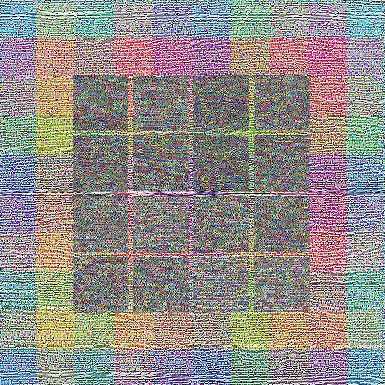
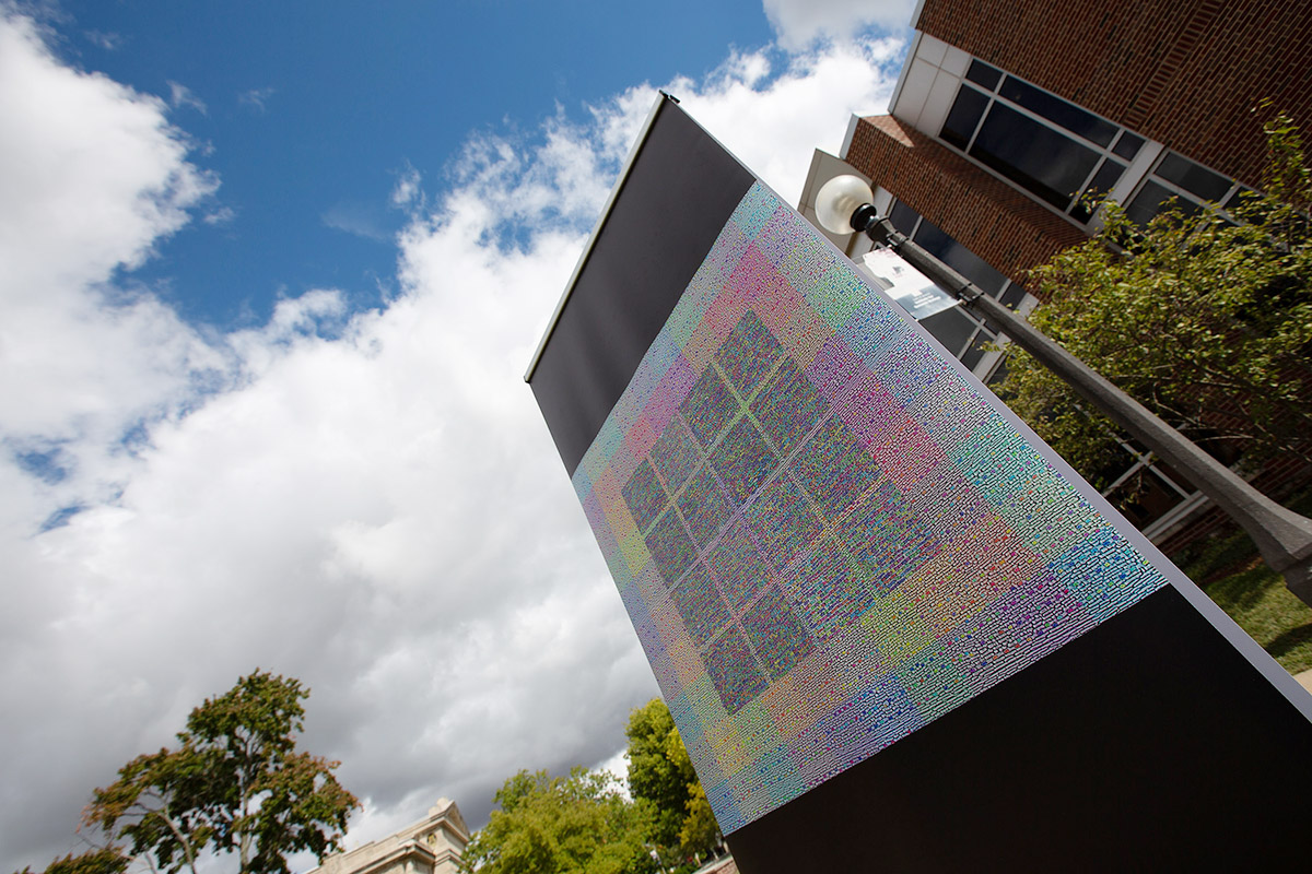
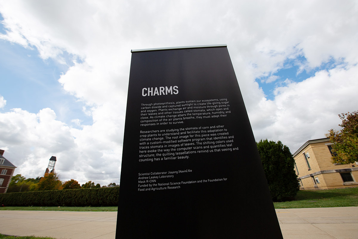
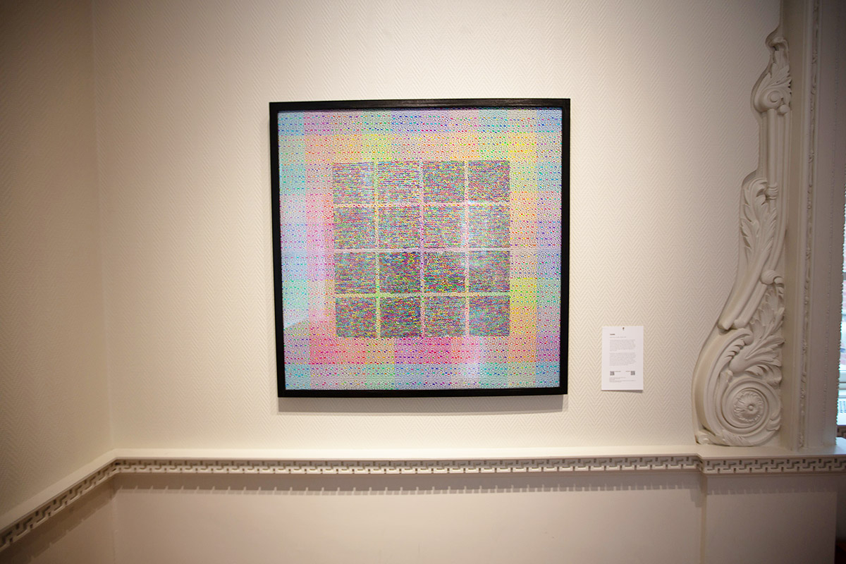
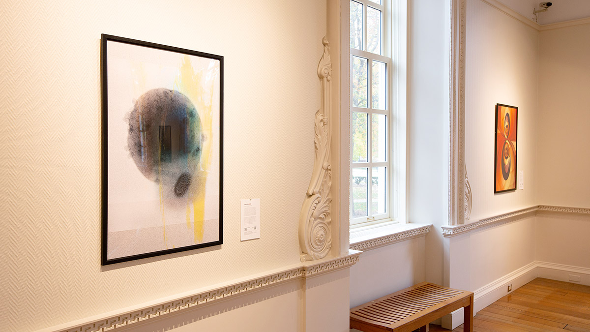
Through photosynthesis, plants sustain our ecosystems, using carbon dioxide and captured sunlight to create life-giving sugar and oxygen. Plants exchange air and moisture through pores in their leaves and other tissues called stomata, which open and close. As climate change alters the temperature, humidity, and composition of the air plants breathe, they must adapt their responses in order to survive.
Researchers are studying the stomata of corn and other crop plants to understand and facilitate this adaptation to climate change. The root image for this piece was created with a custom-modified software program that identifies and traces stomata in images of leaves. The shifting colors used here evoke the way the computer scans and quantifies leaf structure; the quilting tessellations remind us that seeing and counting has a familiar beauty.
To enjoy the show remotely, view the works and accompanying videos below. Select pieces include brief discussions between the artist and the scientist about the featured work.
Life in the Dunes
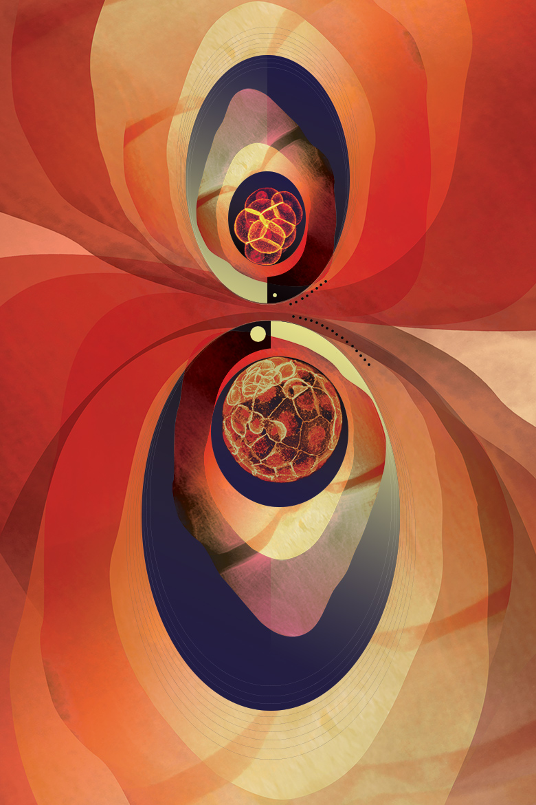
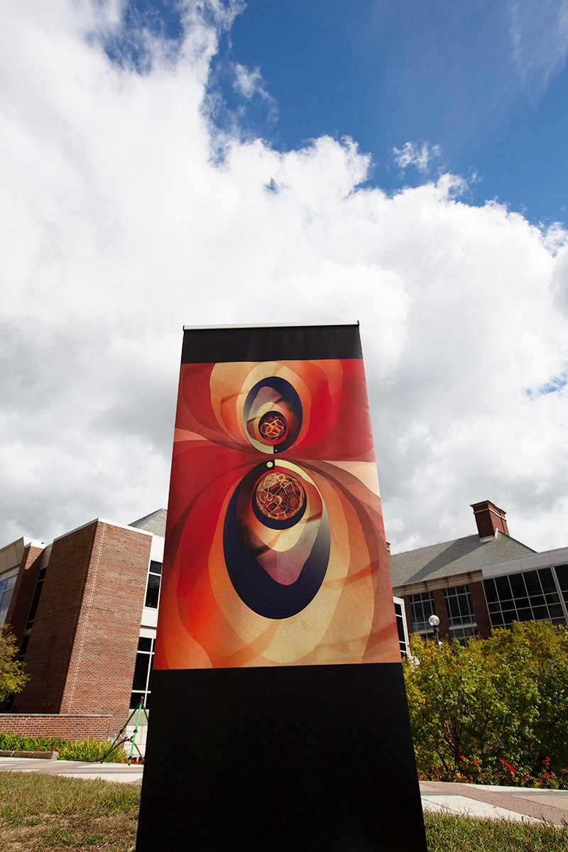
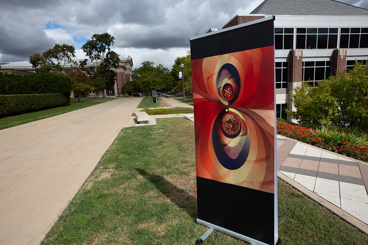
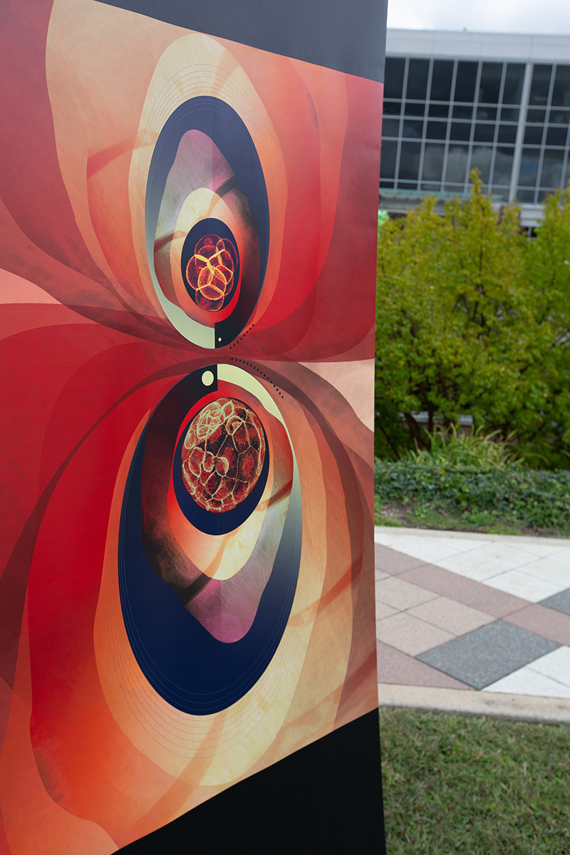
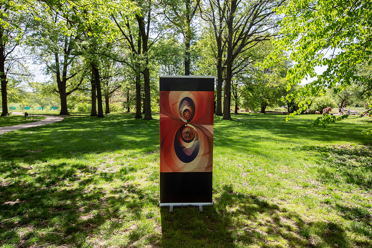
Embryonic development, or embryogenesis, represents the crucial early stages of prenatal development where the embryo forms and begins to take shape. Among other factors, the environment can play a role in the pregnancy and in turn, development of the embryo. Researchers are investigating the impact of the pregnant person’s exposure to environmental toxicants during the early stages of pregnancy.
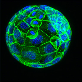
Shown here is a two day-old embryo (top image) and a five day-old blastocyst (bottom image) from mice taken at 20X magnification. The images are adorned with layers of CT scans of uteruses and knee joints and small dots representing the number of cells in each image. Through its soft tones and robust images, this piece reminds us of the delicate yet powerful nature of reproduction.
To enjoy the show remotely, view the works and accompanying videos below. Select pieces include brief discussions between the artist and the scientist about the featured work.
Heated Battleground
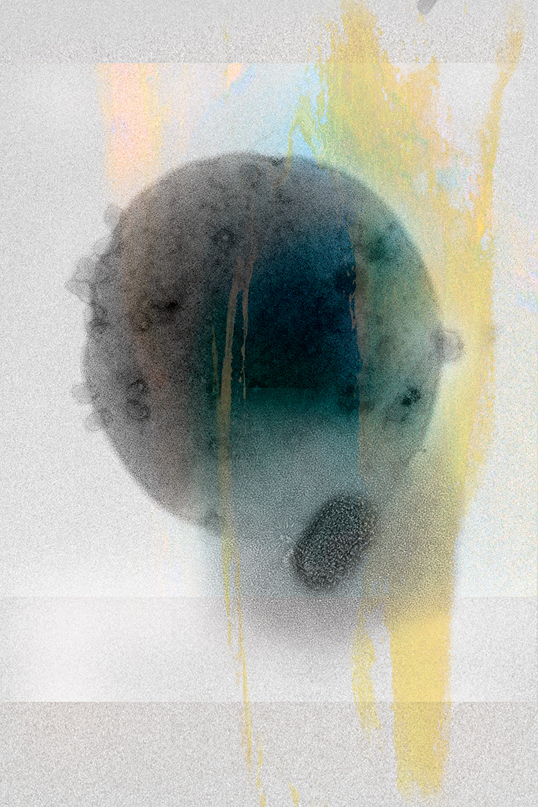
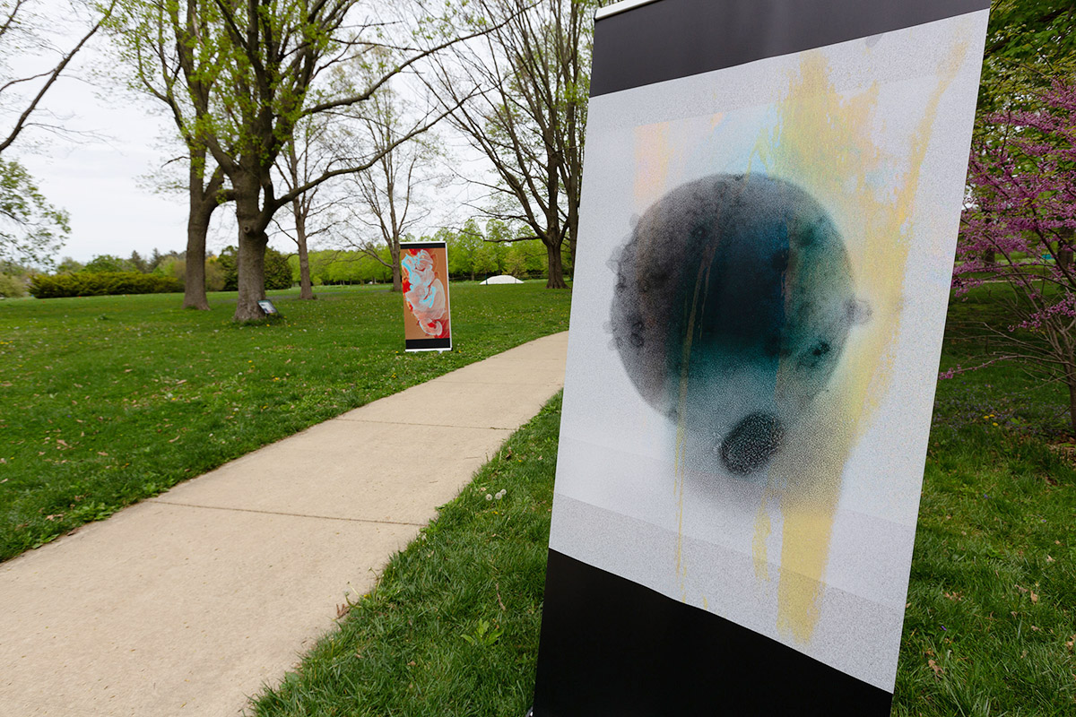
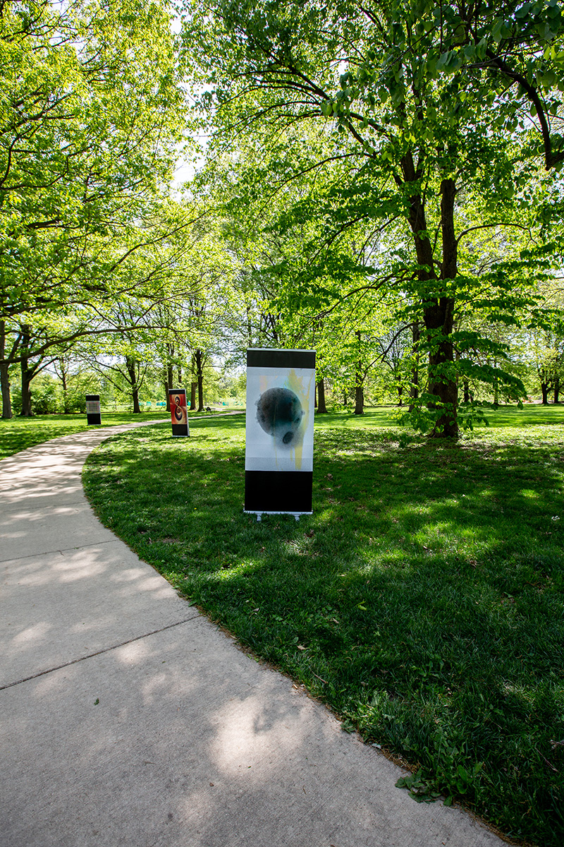
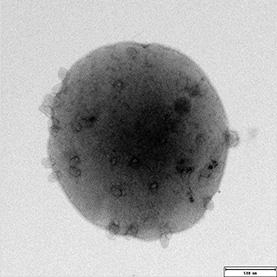
Viruses are capable of infecting every single life form on Earth, with the number of viral particles exceeding the number of stars in the universe. The archaeon Sulfolobus islandicus, which can be isolated from hot springs, was found to be infected by a novel virus called Sulfolobus super-elliptical virus (SSeV). By examining virus-host interactions in a closed system, researchers can understand ecological dynamics within a population, having a broader impact on human health applications.
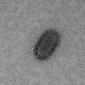
These transmission electron microscopy (TEM) images show SSeV and viral progeny budding from the Sulfolobus islandicus host. The vibrant hues reflect the geothermal pools that serve as the battleground for Sulfolobus islandicus and its viruses. This piece captures the ethereal beauty of a simple system while revealing the dynamic interactions between a host and its virus.
To enjoy the show remotely, view the works and accompanying videos below. Select pieces include brief discussions between the artist and the scientist about the featured work.
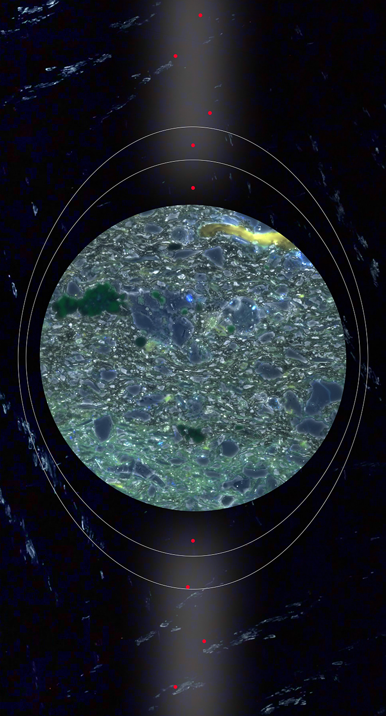
From the Depths
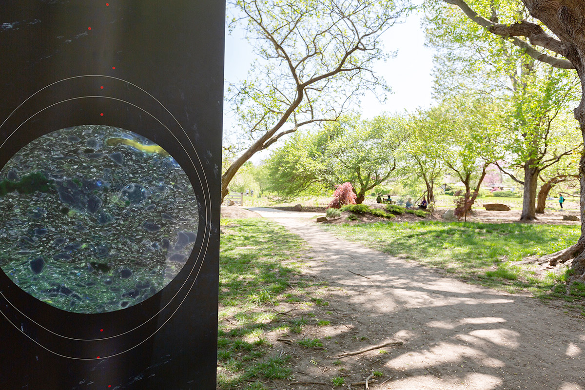
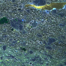
The constant demand for oil and gas has dramatically increased hydraulic fracturing (fracking) of shale reservoirs, decreasing reservoir porosity and permeability. As a result, the control and prevention of bioclogging is important in sustaining hydrocarbon production from fracked shale reservoirs. Using the first-ever constructed shale—GeoBioCell—as a microfluidic test bed, researchers are investigating the effects of sulfate-reducing bacteria and iron sulfide biominerals on porosity and hydraulic resistance in fracked shale reservoirs.
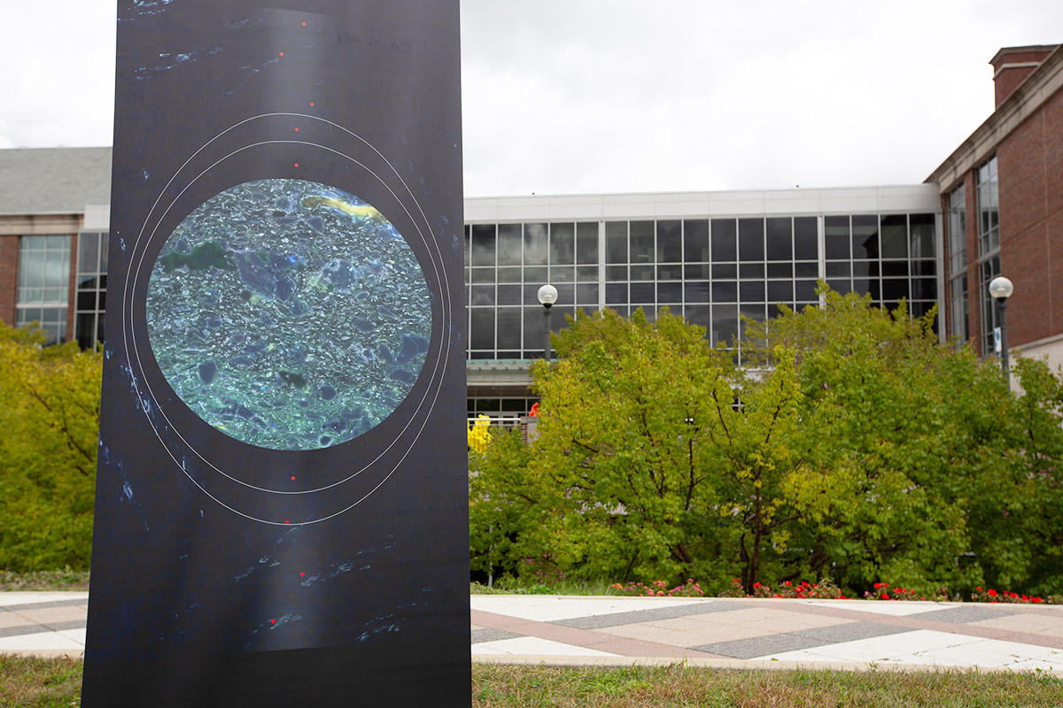
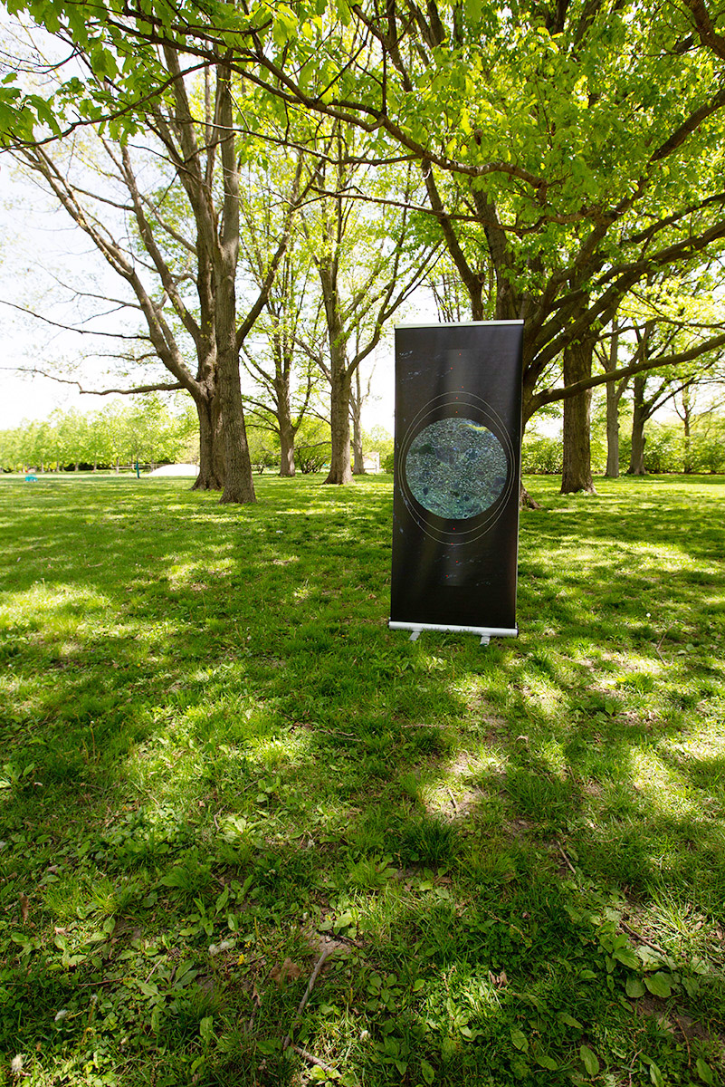
This image of a Devonian-age New Albany shale sample collected from deep within the Illinois Basin was created using Zeiss AxioObserver Z1 Widefield Fluorescence Microscope with Apotome. The light-green and periwinkle blue quartz grains are interspersed with dark-green pyrite, reminiscent of a sea of glass. The dark backdrop accentuates the natural beauty of shale and its formation in ancient sea beds from deep water marine environments.
To enjoy the show remotely, view the works and accompanying videos below. Select pieces include brief discussions between the artist and the scientist about the featured work.
Waves
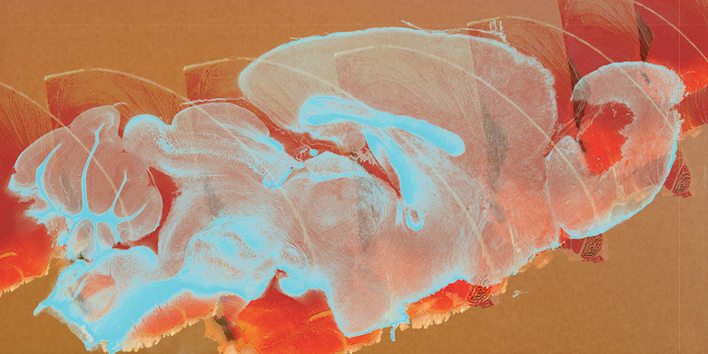
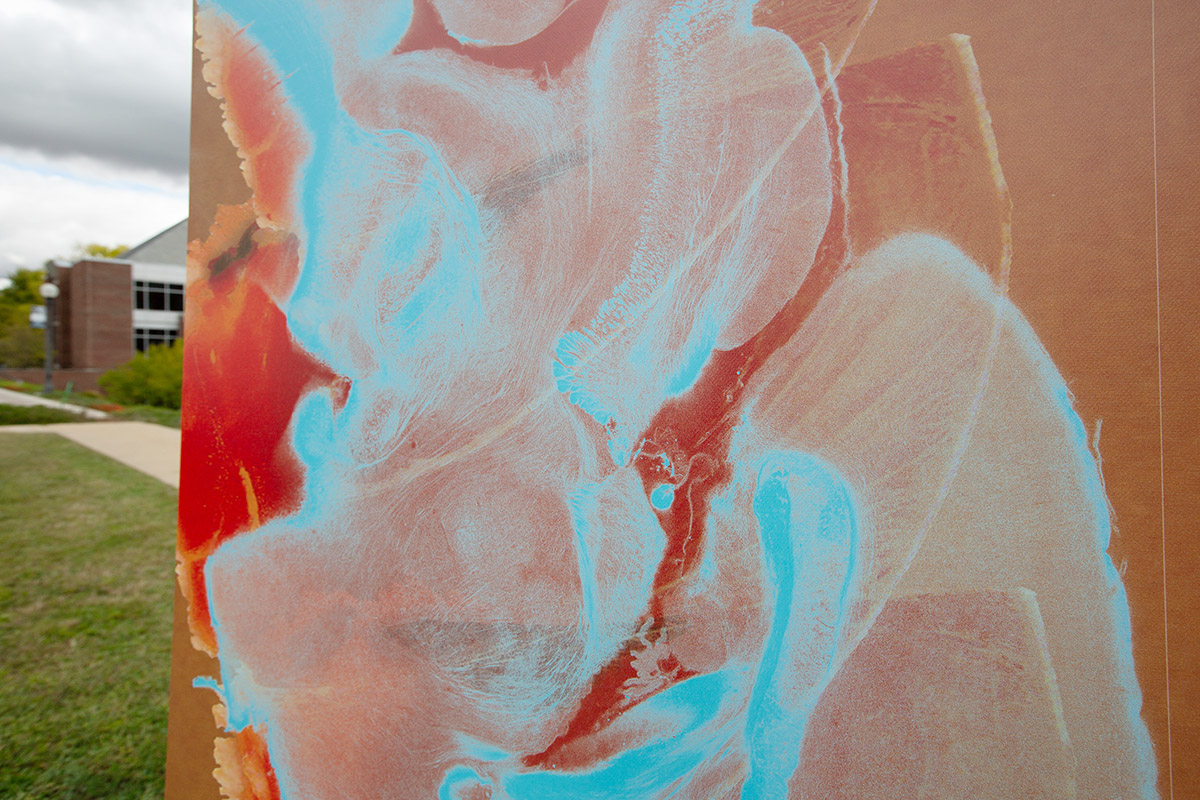
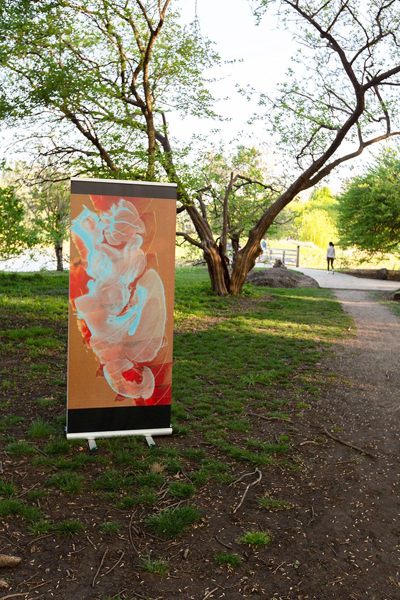
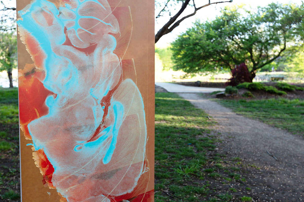
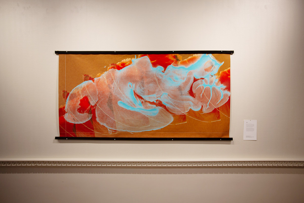
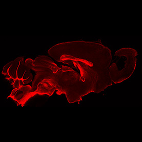
Myelination is an important process in the brain where a fatty substance called myelin is formed around axons of neurons. The formation of myelin increases the speed of electrical signals traveling along the axons, thus playing a vital role in the central nervous system. Researchers are investigating the effects of diet and disease on myelination processes using mouse brains.
A tissue-clearing technique called CLARITY (Clear Lipid-exchanged Acrylamide-hybridized Rigid Imaging/ Immunostainingcompatible Tissue-hYdrogel) was used to render the brain tissue transparent while maintaining an intact protein scaffold. The image, taken with a confocal microscope, shows a sagittal section of the mouse brain immunostained with anti-proteolipid protein. This piece conveys the fragile yet complex nature of the brain.
Scientist Collaborator
Jiayang (Kevin) Xie
Andrew Leakey Laboratory
Instrument
Mask R-CNN
Funding Agency
National Science Foundation
Foundation for Food and Agriculture Research
Original Imaging

Special Thanks
Kleinmuntz Center for Genomics in Business and Society; Nelson family and BodyWork Associates
Image Rights
Images not for public use without permission from the Carl R. Woese Institute for Genomic Biology
Scientist Collaborator
Kadeem A. Richardson
Romana Nowak Laboratory
Instrument
Zeiss LSM 880 Confocal Microscope with Airyscan Superresoution
Funding Agency
National Institutes of Health
Original Imaging
Special Thanks
Kleinmuntz Center for Genomics in Business and Society; Nelson family and BodyWork Associates
Image Rights
Images not for public use without permission from the Carl R. Woese Institute for Genomic Biology
Scientist Collaborator
Elizabeth Rowland
Rachel Whitaker Laboratory
Instrument
Philips CM200 Transmission Electron Microscope
Funding Agency
Space Administration
Mame Shiao Debbie award, Department of Microbiology
Original Imaging
Special Thanks
Kleinmuntz Center for Genomics in Business and Society; Nelson family and BodyWork Associates
Image Rights
Images not for public use without permission from the Carl R. Woese Institute for Genomic Biology
From the Depths
Scientist Collaborators
Bruce W. Fouke and Mayandi Sivaguru
Bruce Fouke Laboratory
Instrument
Zeiss AxioObserver Z1 Widefield Fluorescence Microscope with Apotome
Funding Agency
DuPont Microbial Control
Original Imaging
Special Thanks
Kleinmuntz Center for Genomics in Business and Society; Nelson family and BodyWork Associates
Image Rights
Images not for public use without permission from the Carl R. Woese Institute for Genomic Biology
Scientist Collaborator
Allison Yukiko Louie
Andrew Steelman Laboratory
Instrument
Zeiss 710 Multiphoton Confocal Microscope
Funding Agency
United States Department of Agriculture
University of Illinois at Urbana–Champaign start-up funds
National Multiple Sclerosis Society Pilot Research Grant
Original Imaging
Special Thanks
Kleinmuntz Center for Genomics in Business and Society; Nelson family and BodyWork Associates
Image Rights
Images not for public use without permission from the Carl R. Woese Institute for Genomic Biology
Special Thanks
Champaign businessman Doug Nelson, President of BodyWork Associates, first proposed the idea that became Art of Science, and his continued efforts to support the exhibit made its realization possible. The IGB is also grateful to James Barham of Barham Benefit Group and [co][lab] founder Matt Cho for hosting the annual exhibit.


