CURATED BY
Julia Pollack
WHEN
December 28 - December 28
WHERE
Cafeteria & Company

View Gallery
1
/
10
1 / x

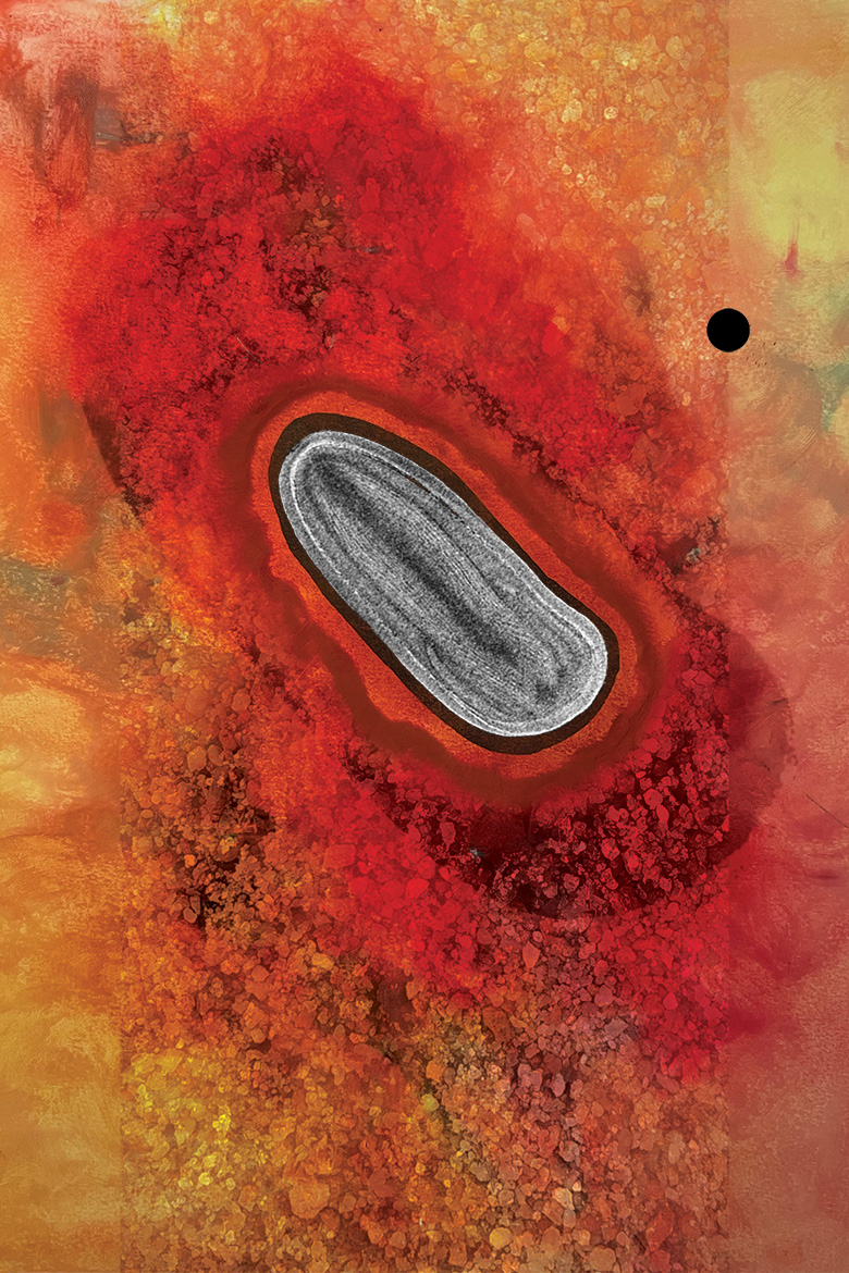
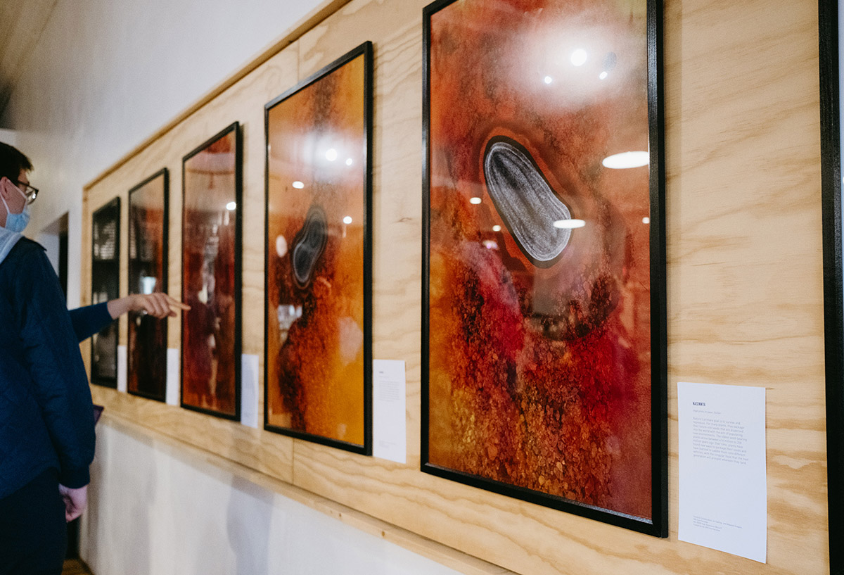
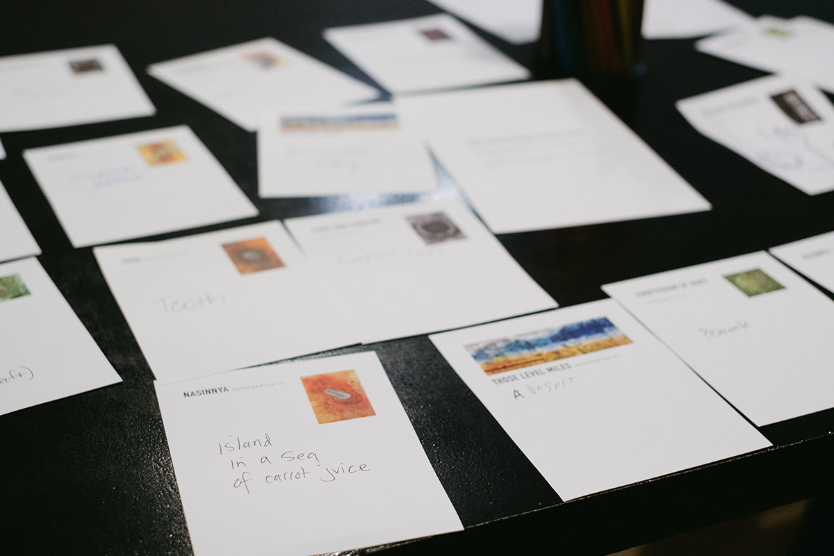
Nasinnya
Nature’s primary goal is to survive and reproduce. For many plants, they package their future into seeds that are dispersed into the world with the aim of populating new environments. The oldest seed-bearing plants arose between 416 million to 358 million years ago. Over time, plants have found new ways to package their seeds and have learned to saddle them onto different vehicles, with the singular hope that the next generation will prosper wherever they land.
Prasnuta
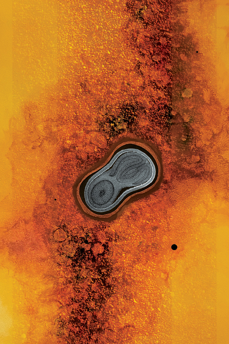
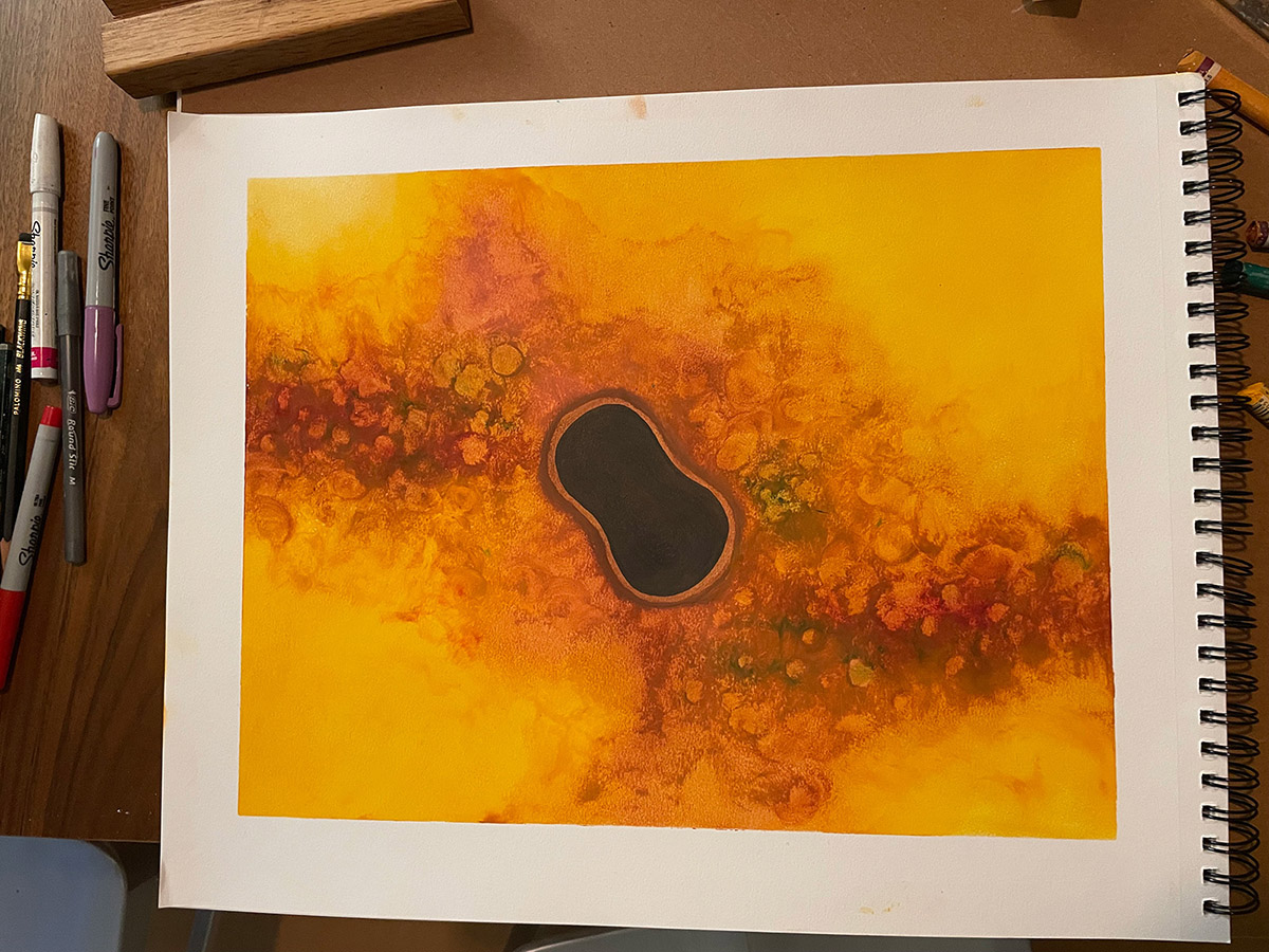
How do plants equip their seeds so that they can succeed in the big bad world? Although the finer details of different seeds vary, they consist of three primary parts. First, they have the embryo, a young multicellular organism which emerges from the seed. Second, seeds contain a source of stored food called the endosperm. And finally, they have protective outer coverings to shield the embryo until it is ready to come out.

Sediz
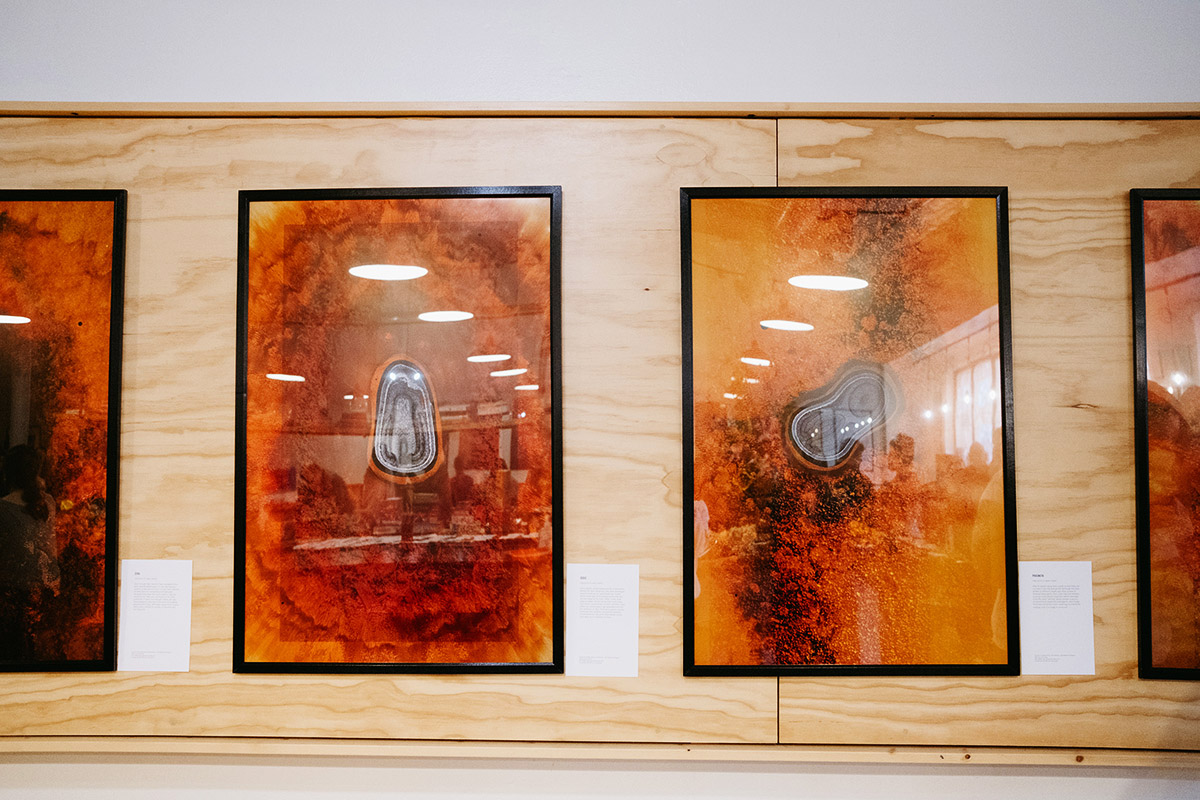
Since plants cannot walk around to find better homes for their offspring, they have developed several methods for seed dispersal. Some plants have light and feathery seeds that are carried away by the wind. Other seeds float away on water, protected by their hard coats. Still others use animals as their vehicles—they either are consumed or get attached onto the fur, feathers, or skin. And some plants choose methods that could be considered violently unconventional, like pine trees whose cones only open up in response to fires.
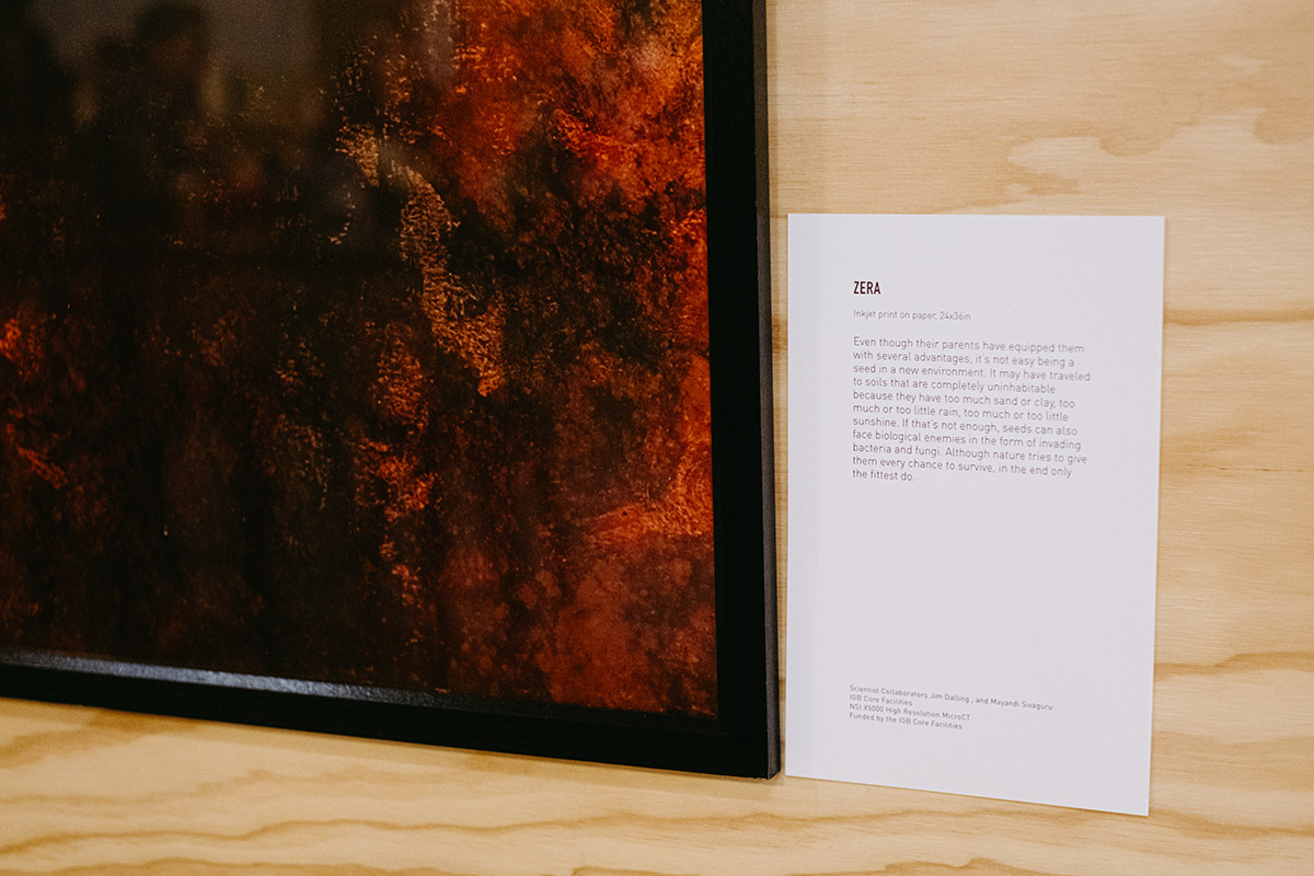
Zera
Even though their parents have equipped them with several advantages, it’s not easy being a seed in a new environment. It may have traveled to soils that are completely uninhabitable because they have too much sand or clay, too much or too little rain, too much or too little sunshine. If that’s not enough, seeds can also face biological enemies in the form of invading bacteria and fungi. Although nature tries to give them every chance to survive, in the end only the fittest do.
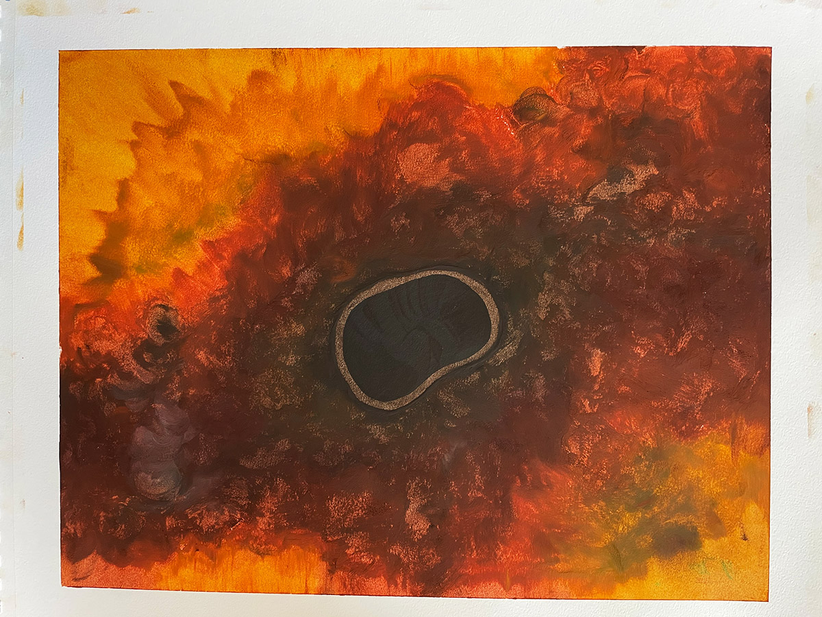
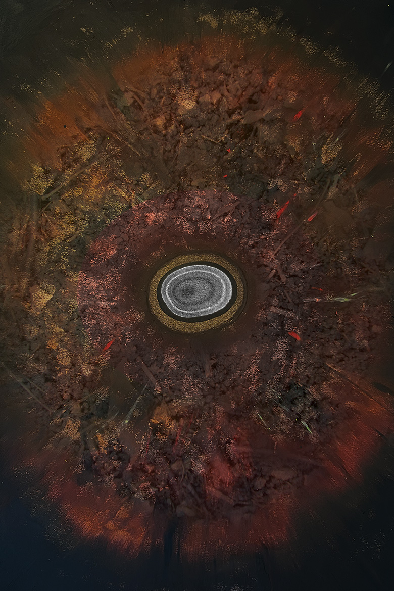
Mokosh's Cave
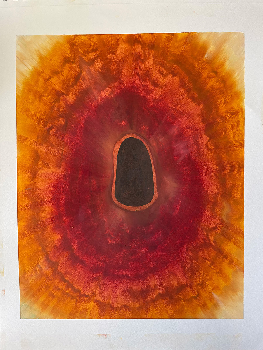
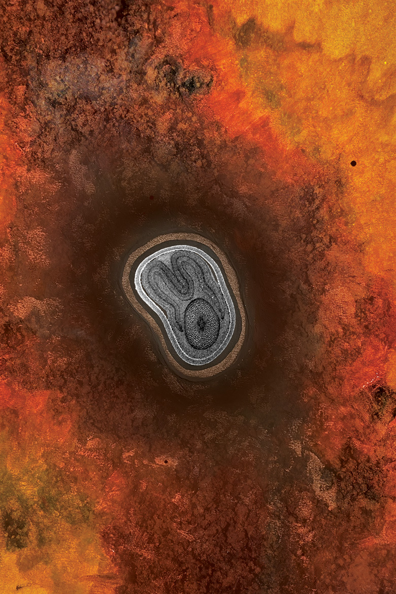
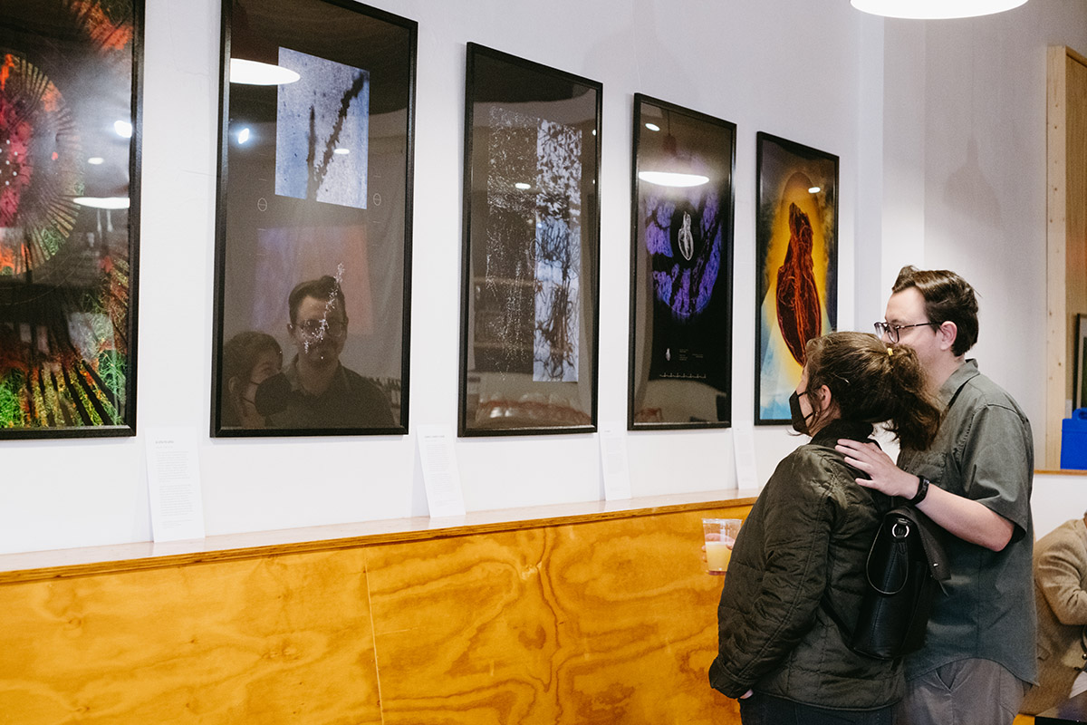
Made with layers of pastel drawings and textural soil images, these five pieces represent a fresh seed lounging in its bed of nutrients. The original images are 2D renderings of 3D images of velvet leaf seeds. The intact seeds have been scanned to see how beneficial fungi can affect their growth.

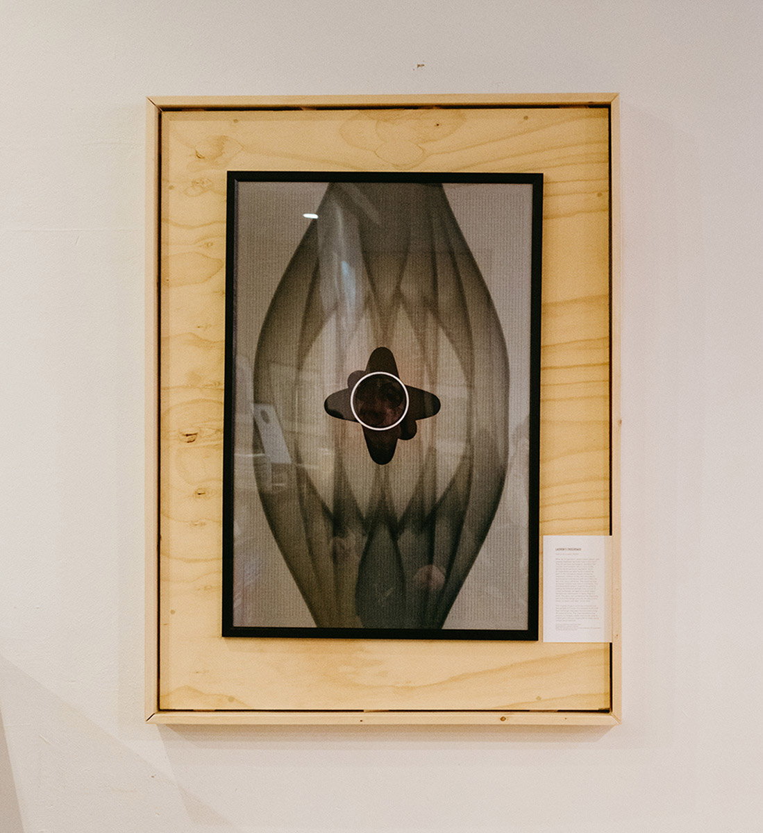
Lasiren's Crossroads
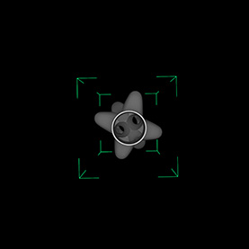
What do refrigerators, steam power plants, and sewage treatment facilities have in common? They all use heat exchangers—systems that transfer heat between two or more fluids, optimizing temperature regulation. Such arrangements are also found in nature as seen in the circulatory system of marine mammals. Arteries to the skin carry warm blood and are intertwined with veins from the skin that carry cold blood. The resulting heat exchange reduces heat loss in cold water. This interdependent system inspired the artist to overlay two different perspectives: a scan of a heat exchanger set against a graph paper background, reminiscent of Agnes Martin’s work. The piece also symbolizes the beginning and end of the design process. An idea is first drawn on paper and later comes alive.
The original image is an X-ray computerized tomography (CT) scan of a compact multi-tube heat exchanger that was manufactured using 3D metal printing. This new manufacturing process is cheaper, easier, and can be used to create parts that are impossible to make using conventional methods.
Accords
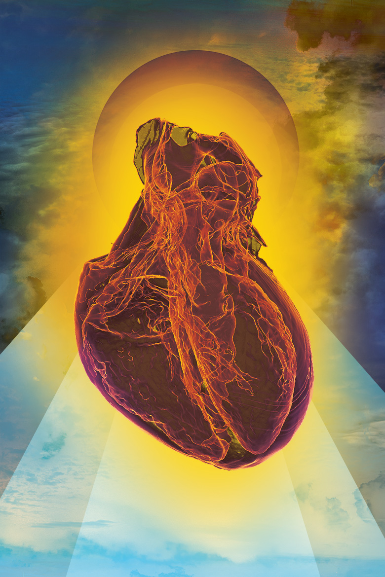
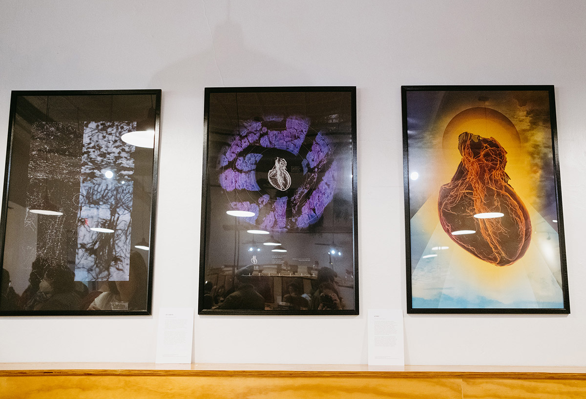
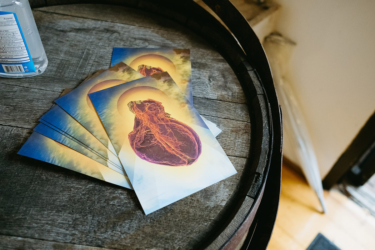
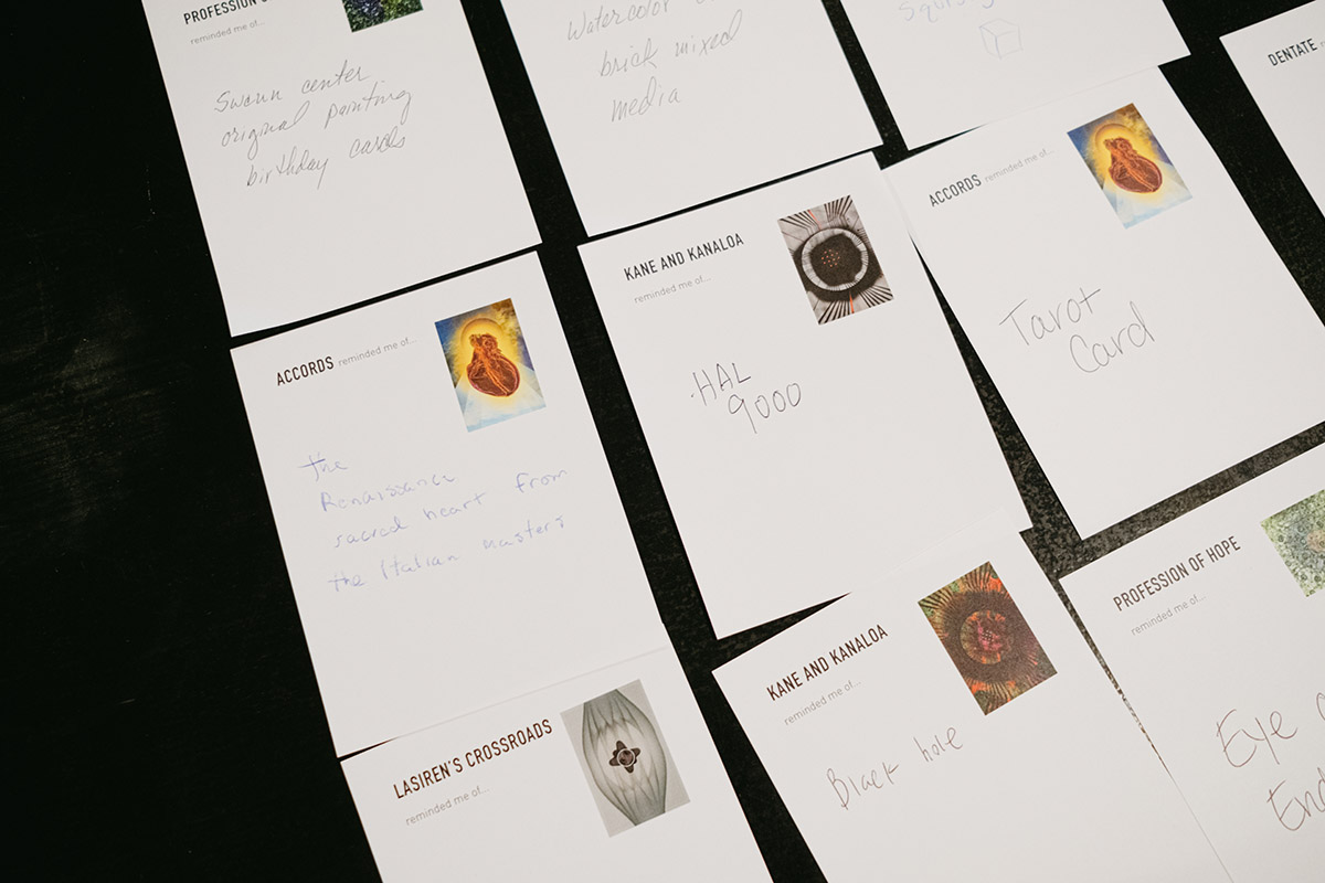
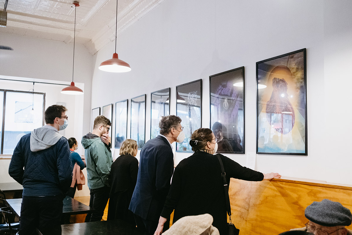
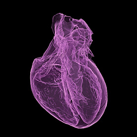
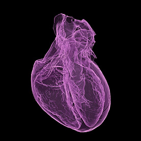
For centuries, the heart has been revered across cultures as the body’s soul and a center for life and love. For example, in Egyptian mythology, the heart was weighed in the afterlife as a measure of morality. The image of the heart also made its way into theology, often bathed in ethereal light to represent love and sacrifice. In this piece, the background has been stitched together from photographs of sunsets. The combination of the heart and sky reference the belief of the heart’s divinity.
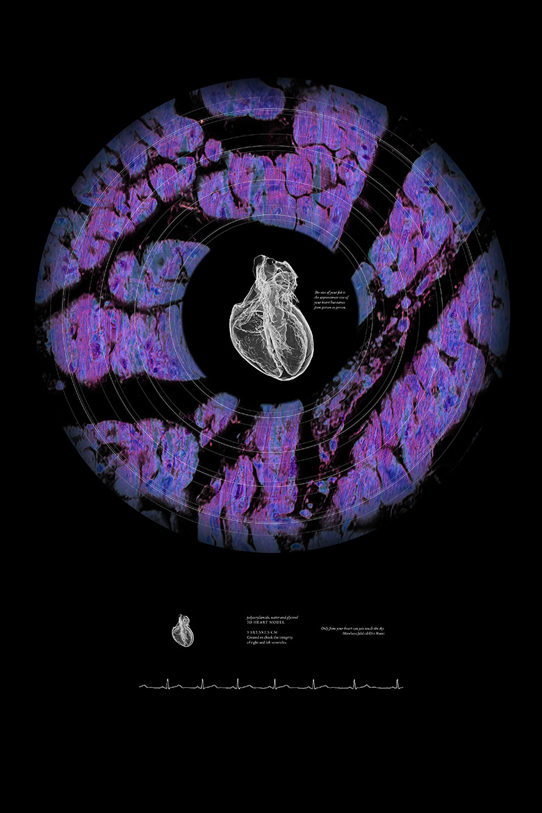
The original image is a heart model that was made by 3D printing a non-drying hydrogel, a gelatin-like material. The prototype, measuring approximately 3.5x2.5x2.5 cm, was created to check the integrity of chambers in the heart. Eventually, a larger model will be printed and used as a tool to help surgeons practice their technique before they operate on patients.
Joy's Bonfire
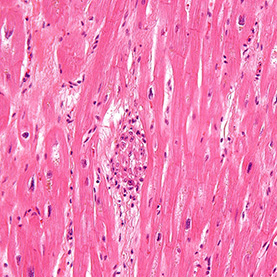
Our hearts are constantly receiving and pumping out blood, generally heard as a “lub-dub” sound through a stethoscope. The sound occurs when the heart muscle relaxes and refills with blood and when it contracts to send blood back. A healthy heart takes about 0.8 seconds to complete the cycle, resulting in a typical rate of 70 to 75 beats per minute.
Amazingly, heartbeats can synchronize when people are captivated by the same story, sleeping next to each other, or participating in religious rituals such as dhikr in Sufism. Often referred to as “the way of the heart,” the ritual involves deep meditations, ecstatic trances, and sacred melodies and movements, including spinning the body in repetitive circles. Researchers have shown that the Sufis’ quest to unify hearts in celebration of God goes beyond metaphor: Their hearts really do beat as one.
Dimensional Transcendentalism
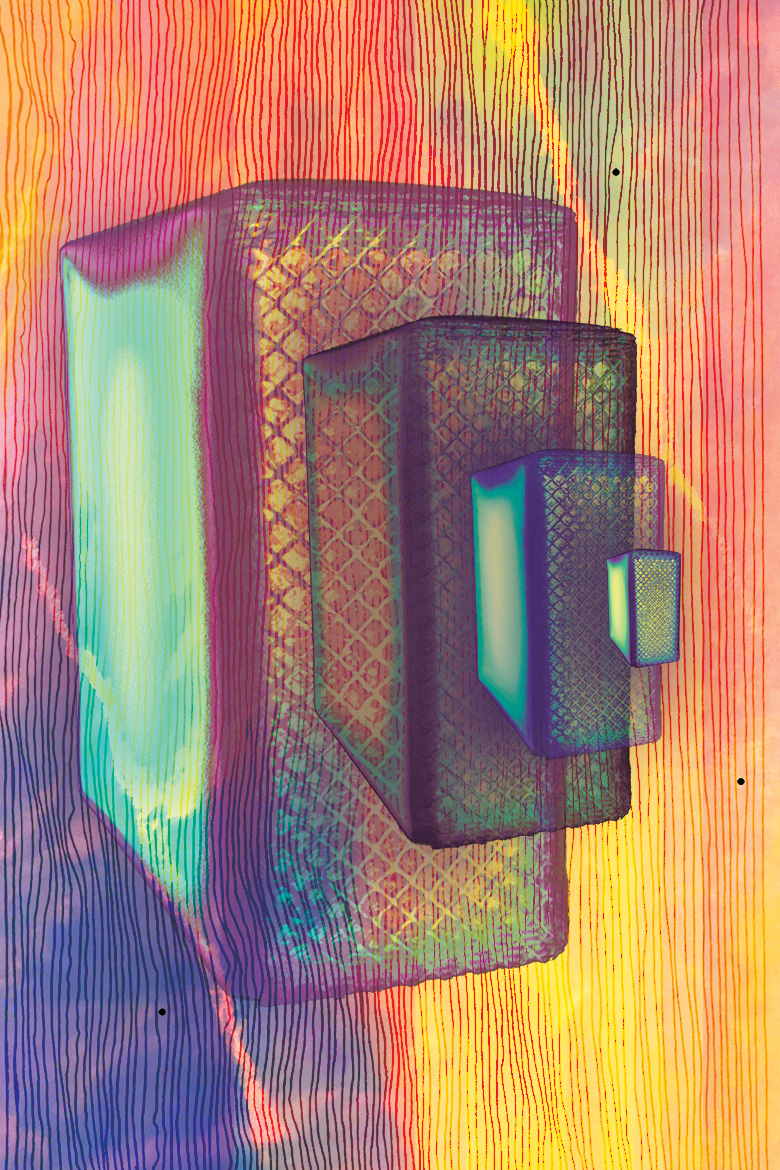
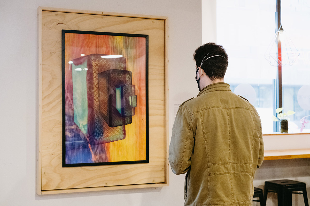
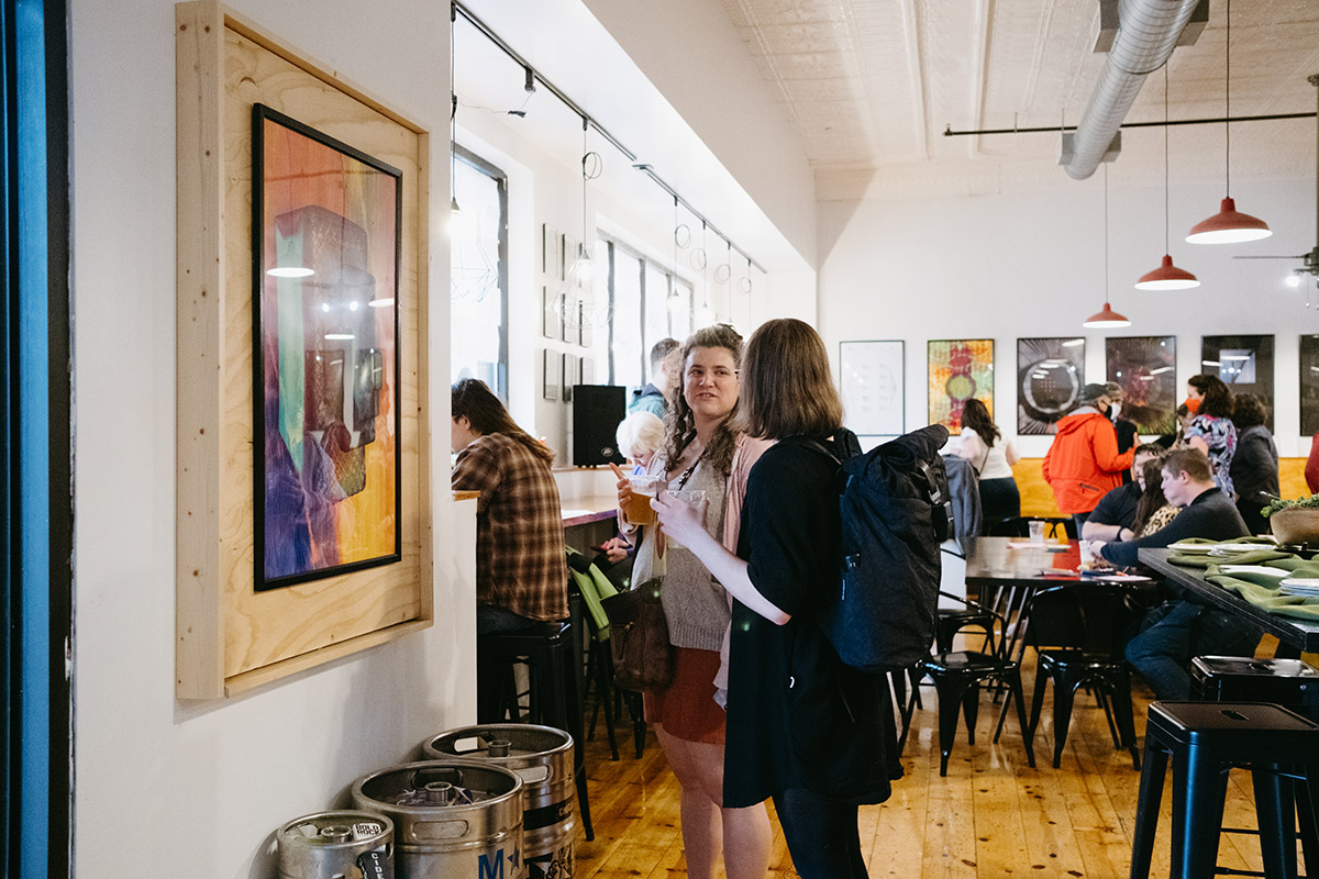
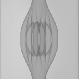
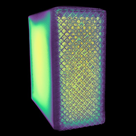
A camera requires two opposite but interconnected facets: light and dark. It is a sealed box with a small hole that allows light to pass through onto a light-sensitive surface. Too much light causes a washed out photo and too little captures nothing. The piece evokes 19th century cameras, which had accordion-like bellows that helped the photographer capture objects from close quarters. The artist included her own drawings to add color and texture to the piece. The bricks have been layered with different opacities and set against a background of sunset images. The result mirrors our reaction to beautiful skies—a feeling of awe followed by a quick photo.
The original image is a computerized tomography (CT) scan of a stainless steel block that was manufactured by a 3D-printing process. The scan helps engineers determine defects produced in the material during the manufacturing process.

Kane and Kanaola
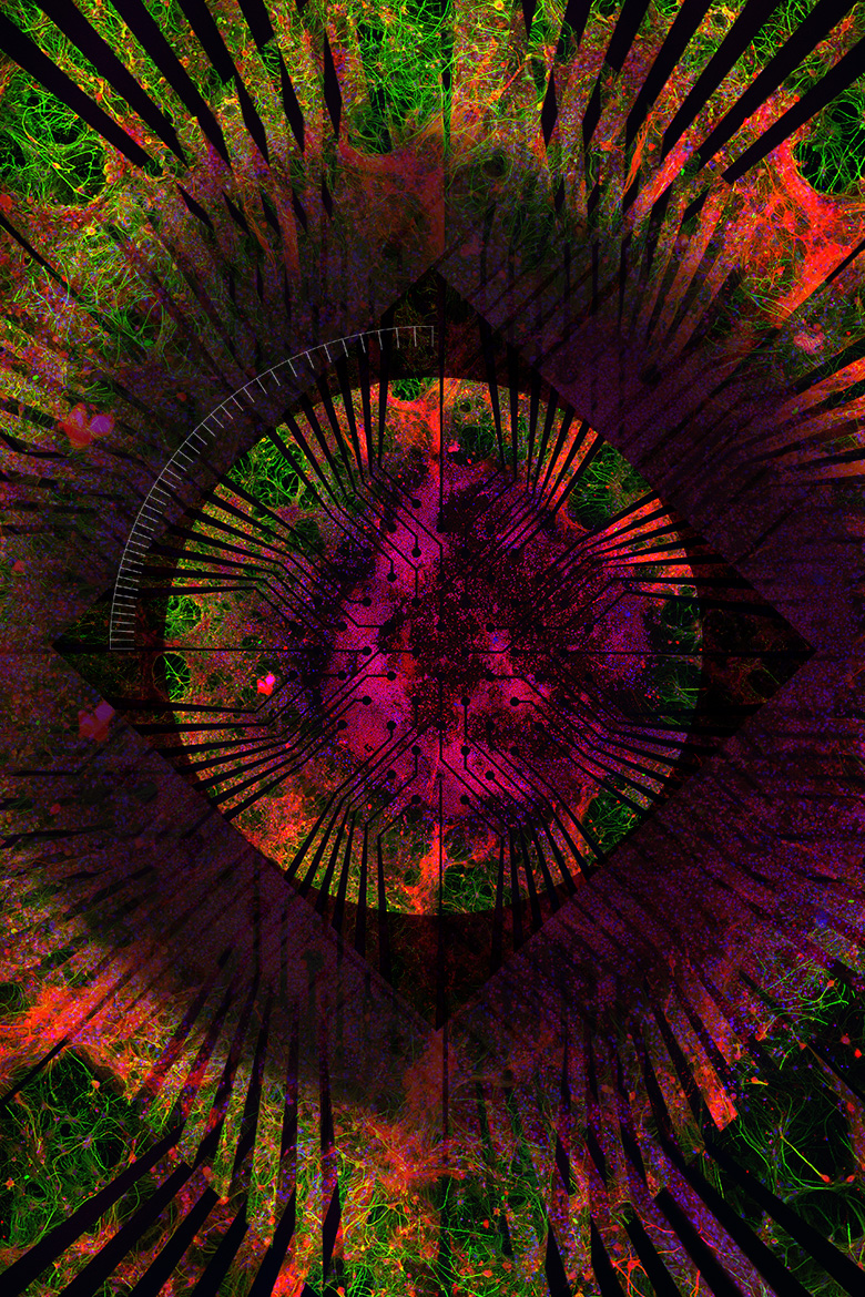
When we think of robots, we often picture rigid Terminator-style giants with halting, awkward gaits. While miniature versions of these exist, usually made of metals, ceramics, and hard plastics, there has also been a push to develop soft robots that take on tasks that hard robots cannot. Inspired by soft creatures, like octopi that have entirely pliable bodies, these machines can help in invasive surgery or disaster relief scenarios, where they need to squeeze through small crevices.
The original research image depicts a substrate that is being developed for soft robotics to help researchers study the interface between nerves and muscle fibers. Created by layering the brain tissue with the substrate, the composition reflects the back-and-forth communication that occurs between us and the gadgets we control.
Kane and Kanaloa
When we think of robots, we often picture rigid Terminator-style giants with halting, awkward gaits. While miniature versions of these exist, usually made of metals, ceramics, and hard plastics, there has also been a push to develop soft robots that take on tasks that hard robots cannot. Inspired by soft creatures, like octopi that have entirely pliable bodies, these machines can help in invasive surgery or disaster relief scenarios, where they need to squeeze through small crevices.
The original research image depicts a substrate that is being developed for soft robotics to help researchers study the interface between nerves and muscle fibers. Created by layering the brain tissue with the substrate, the composition reflects the back-and-forth communication that occurs between us and the gadgets we control.
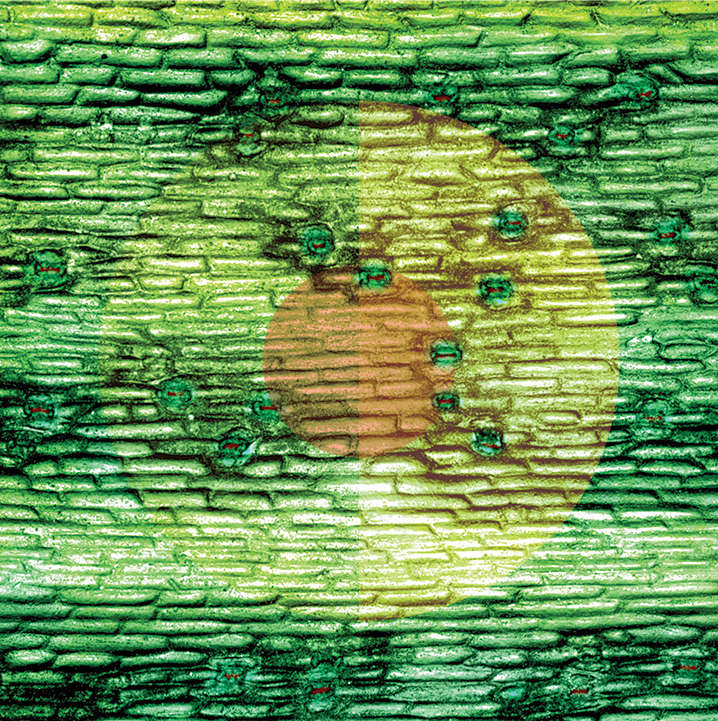
Profession of hope (Ponytail Palm, Beaucarnea recurvata)
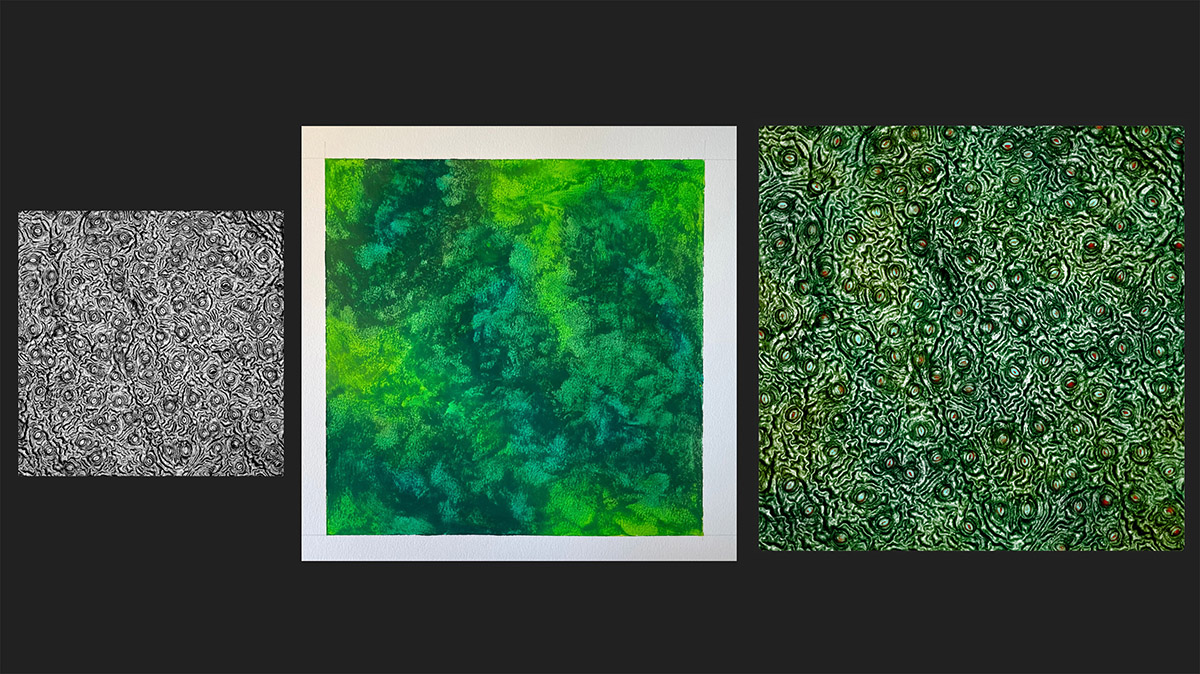
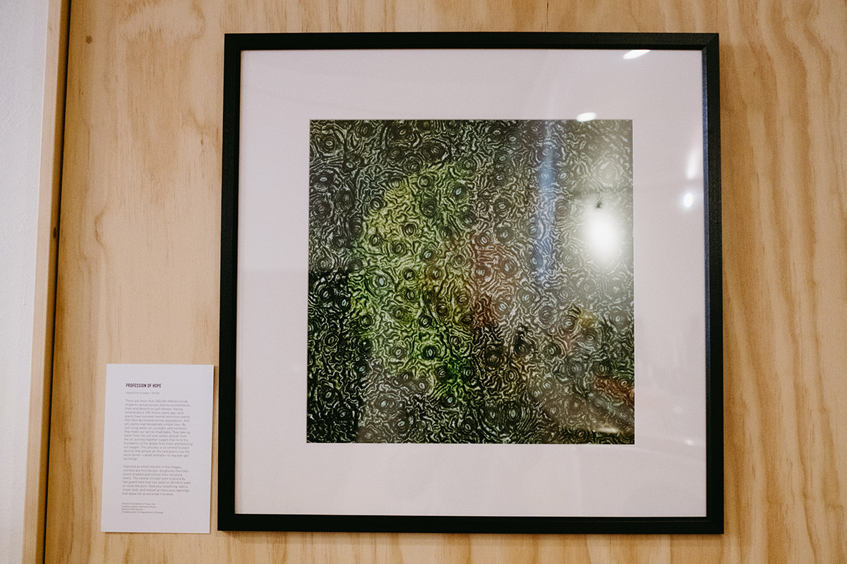
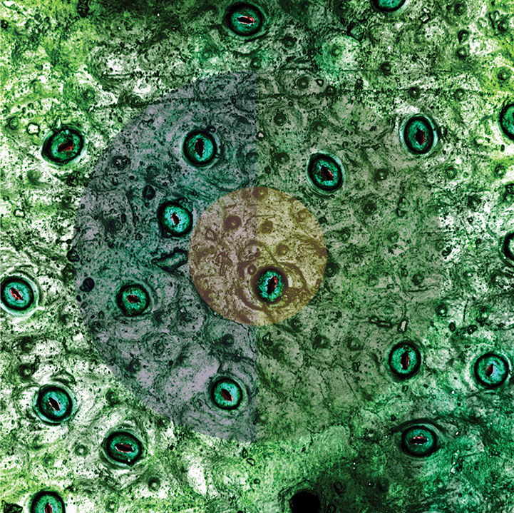
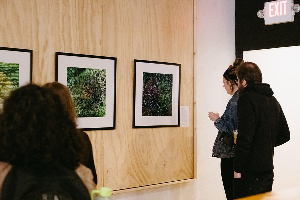
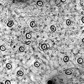
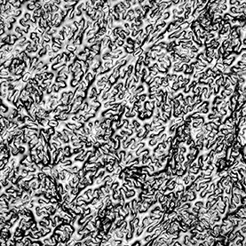
There are more than 300,000 different kinds of plants spread across diverse environments, from arid deserts to lush forests. Having evolved about 700 million years ago, land plants have survived several extinction events that have decimated animal populations. And yet, plants lead deceptively simple lives. By just using water, air, sunlight, and nutrients, they make our world inhabitable. They take up water from the soil and carbon dioxide from the air, putting together sugars that form the foundation of the global food chain and belching out oxygen. This process is so central to plant identity that almost all the land plants use the same pores—called stomata—to regulate gas exchange.
Depicted as small mouths in the images, stomata are microscopic doughnuts that help plants breathe and control their moisture levels. The central circular pore is bound by two guard cells that can swell or shrink to open or close the pore. Slow your breathing, take a closer look, and marvel at innocuous openings that allow life as we know it to exist.
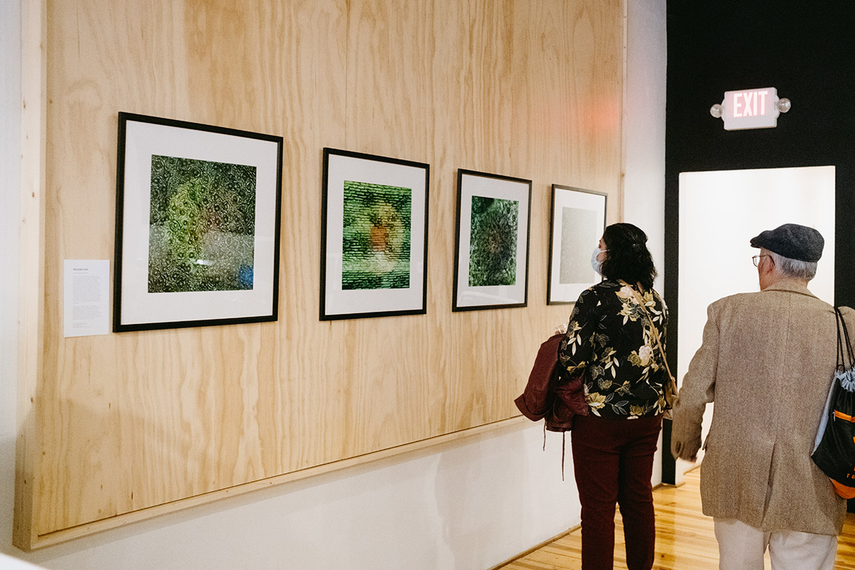
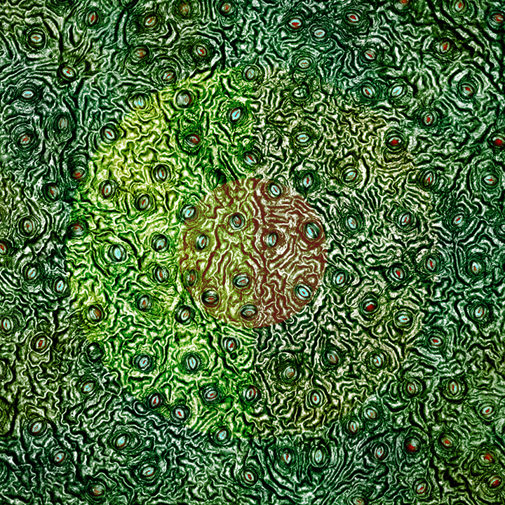
Profession of Hope (Common Ivy, Hedera helix)
There are more than 300,000 different kinds of plants spread across diverse environments, from arid deserts to lush forests. Having evolved about 700 million years ago, land plants have survived several extinction events that have decimated animal populations. And yet, plants lead deceptively simple lives. By just using water, air, sunlight, and nutrients, they make our world inhabitable. They take up water from the soil and carbon dioxide from the air, putting together sugars that form the foundation of the global food chain and belching out oxygen. This process is so central to plant identity that almost all the land plants use the same pores—called stomata—to regulate gas exchange.
Depicted as small mouths in the images, stomata are microscopic doughnuts that help plants breathe and control their moisture levels. The central circular pore is bound by two guard cells that can swell or shrink to open or close the pore. Slow your breathing, take a closer look, and marvel at innocuous openings that allow life as we know it to exist.
Profession of Hope (Wax plant, Hoya carnosa veriegata)
There are more than 300,000 different kinds of plants spread across diverse environments, from arid deserts to lush forests. Having evolved about 700 million years ago, land plants have survived several extinction events that have decimated animal populations. And yet, plants lead deceptively simple lives. By just using water, air, sunlight, and nutrients, they make our world inhabitable. They take up water from the soil and carbon dioxide from the air, putting together sugars that form the foundation of the global food chain and belching out oxygen. This process is so central to plant identity that almost all the land plants use the same pores—called stomata—to regulate gas exchange.
Depicted as small mouths in the images, stomata are microscopic doughnuts that help plants breathe and control their moisture levels. The central circular pore is bound by two guard cells that can swell or shrink to open or close the pore. Slow your breathing, take a closer look, and marvel at innocuous openings that allow life as we know it to exist.
Profession of Hope (Basil, Ocimum basilicum)
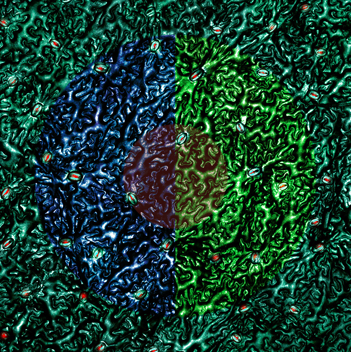
There are more than 300,000 different kinds of plants spread across diverse environments, from arid deserts to lush forests. Having evolved about 700 million years ago, land plants have survived several extinction events that have decimated animal populations. And yet, plants lead deceptively simple lives. By just using water, air, sunlight, and nutrients, they make our world inhabitable. They take up water from the soil and carbon dioxide from the air, putting together sugars that form the foundation of the global food chain and belching out oxygen. This process is so central to plant identity that almost all the land plants use the same pores—called stomata—to regulate gas exchange.
Depicted as small mouths in the images, stomata are microscopic doughnuts that help plants breathe and control their moisture levels. The central circular pore is bound by two guard cells that can swell or shrink to open or close the pore. Slow your breathing, take a closer look, and marvel at innocuous openings that allow life as we know it to exist.
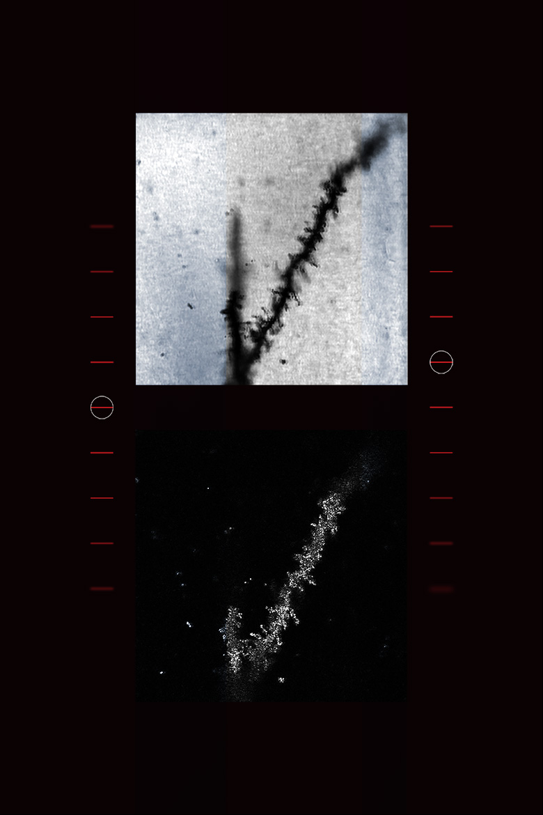
Shadow of a shadow of a shadow
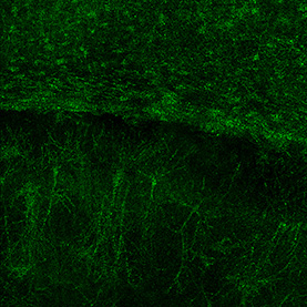
Of all the instruments used in the study of biology, the microscope has contributed the most to furthering knowledge. Surprisingly, it took around 1,300 years from the time optical magnification was discovered utill its first practical application, the invention of eyeglasses in the 13th century. It took another 300 hundred years to invent the microscope. Astoundingly, in the past 430 years we have moved from having microscopes that can give us vague cell outlines to super-resolution microscopes that can be used to track individual molecules inside cells.
This piece is derived from a bright-field microscope image of neurons in the mouse hippocampus: a complex brain structure that has a major role in learning and memory. The technique is the simplest of all optical microscopy approaches. However, its ease also makes it popular, generating images of dark samples against a bright background, allowing scientists to visualize cells across the three domains of life.
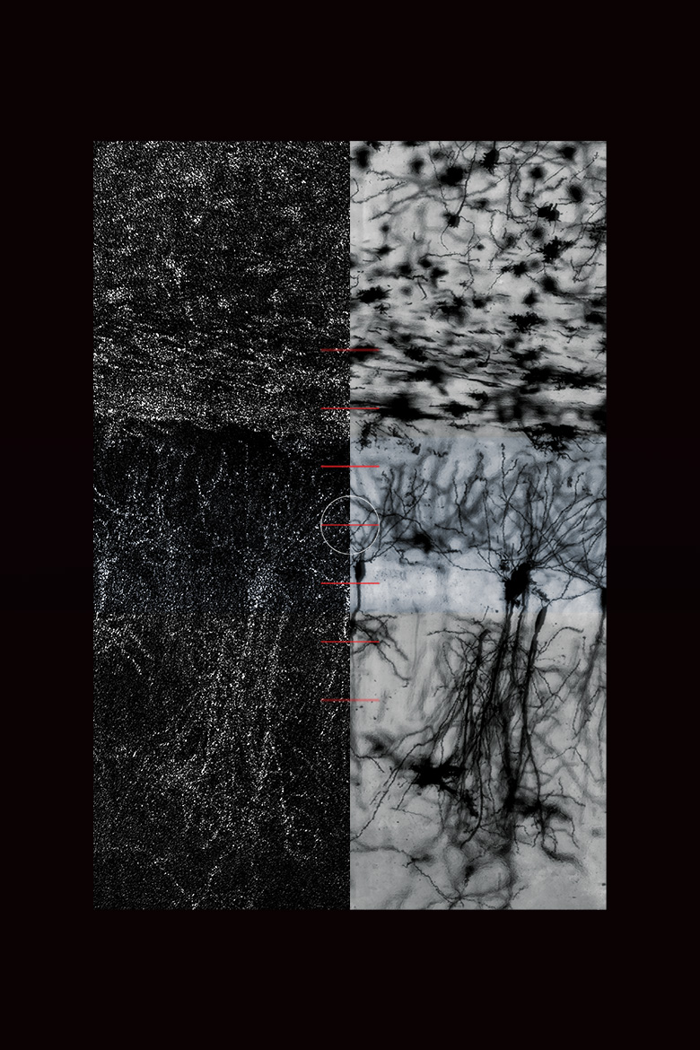
Ad astra per aspera
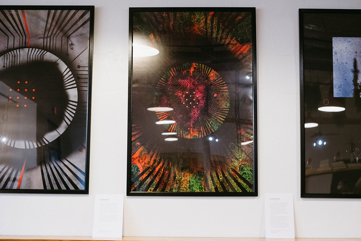
As our understanding of biology has developed, so too have the types of questions we ask. The concept of biology as a single coherent field arose in the 19th century, emerging from traditions of medicine and natural history. Scientists studied what they could observe with their naked eye, making breakthroughs in botany and zoology. After the invention of microscopes, their questions become increasingly detail-oriented: what are cells made up of and how do the molecules interact with each other?
Confocal microscopy, represented in the image, allows us to light up molecules like stars in the night sky. The technique helps scientists observe how cellular components are arranged, creating a picture of zipping molecules in the busy metropolis of a cell. Set up like the abstract tapestries in Barnet Newman paintings, the two sides represent the feeling of a microscope trying to focus from the bigger picture of a mouse hippocampal neuron seen in a bright-field microscope to the inner details brought out by a confocal microscope.
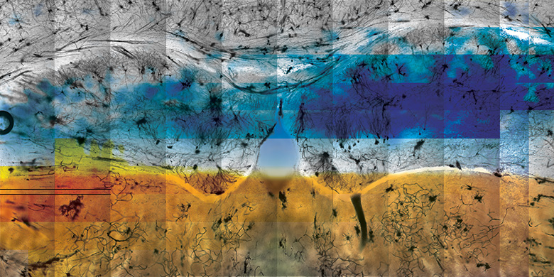
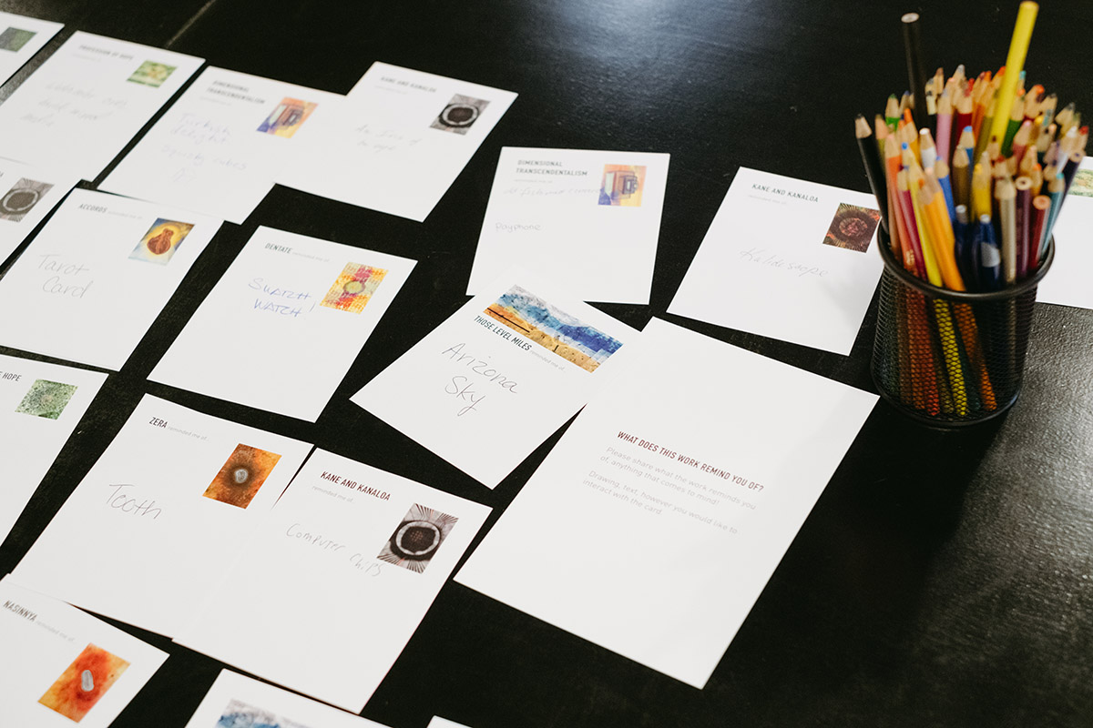
Those Level Miles
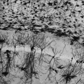
An old tune, the smell of crackling firewood, the sight of your childhood home. All these, and many more, experiences can pull us back into fond memories. What helps us remember? The answer is a seahorse-shaped tube– called the hippocampus–in our brain. Scientists believe that the structure takes simultaneous memories from different sensory regions of the brain and creates a single memory. For example, you may remember driving along the highway instead of having separate memories of the cornfields and the sound of the radio.
In the original image, the researchers stained the hippocampus of mice to study their neuronal processes. The art piece has been created by using a section of the hippocampus and the image of a corn field. It reminds us that our body is constantly changing, forging new memories using a collage of sensations.
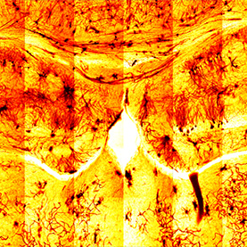
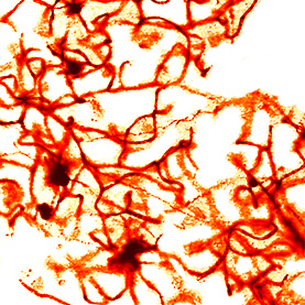
The researchers used Golgi staining to identify structural and organizational differences in neuronal processes of mice with a mutation in an autism susceptibility gene, Auts2.
Flux
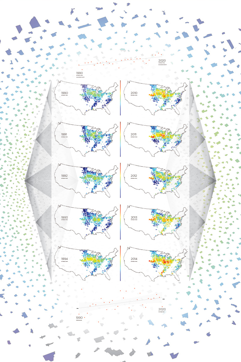
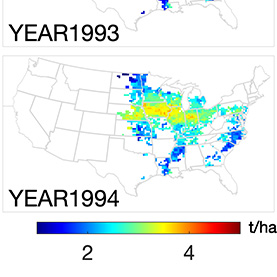
It is often tempting to dwell on what we have failed to achieve: cures for diseases like cancer and Alzheimer’s, eliminating world hunger, or preventing natural disasters. While it is important to remember what needs to be done, it is equally crucial to recognize what we have accomplished so far. In just the past three decades, science has progressed in leaps and bounds with the development of the internet, solar energy devices, antiretroviral treatment for AIDS, and increased agricultural production, among many other advances.
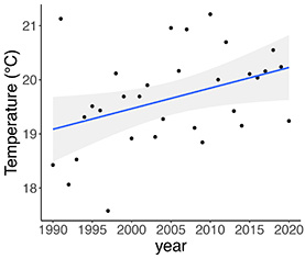
The US is the largest soybean grower in the world, shown in the agricultural productivity maps in this piece. The yield has been changing in both space and time for many decades due to a combination of factors, including technology improvement, climate change, and land use management. Set against a background of US county shapes, the image reminds us that even if we don’t know all the answers, we’re much closer than we were 30 years ago.
Data notes: USDA National Agricultural Statistics Service Information; Water and Atmospheric Resources Monitoring Program; Illinois Climate Network.
Note: The temperature shown are the yearly growing-seaon averages. (i.e., annual mean throughout April-September)
Dentate
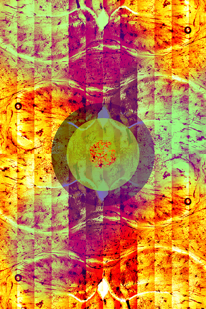
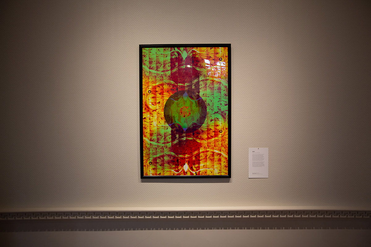
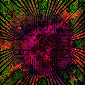
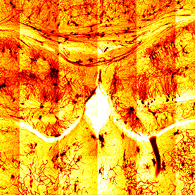
For years, researchers have exploited mice as powerful genetic tools to answer long standing questions in biology. By studying the regulatory networks in the mouse brain, researchers can better understand the link between genomic mechanisms and social behavior. The original image shows a Golgi-stained mouse hippocampus—a region of the brain involved in memory—that was taken with a multiphoton confocal Zeiss 710 microscope. The characteristic black color originates from silver nitrate used in the staining process.
The researchers used Golgi staining to identify structural and organizational differences in neuronal processes of mice with a mutation in an autism susceptibility gene, Auts2. The art piece features a central cluster of neurons, called the dentate nucleus, against a backdrop of psychedelic hippocampus. This piece pays homage to late abstract painter Helen Frankenthaler, who once said to “let the picture lead you where it must go.”
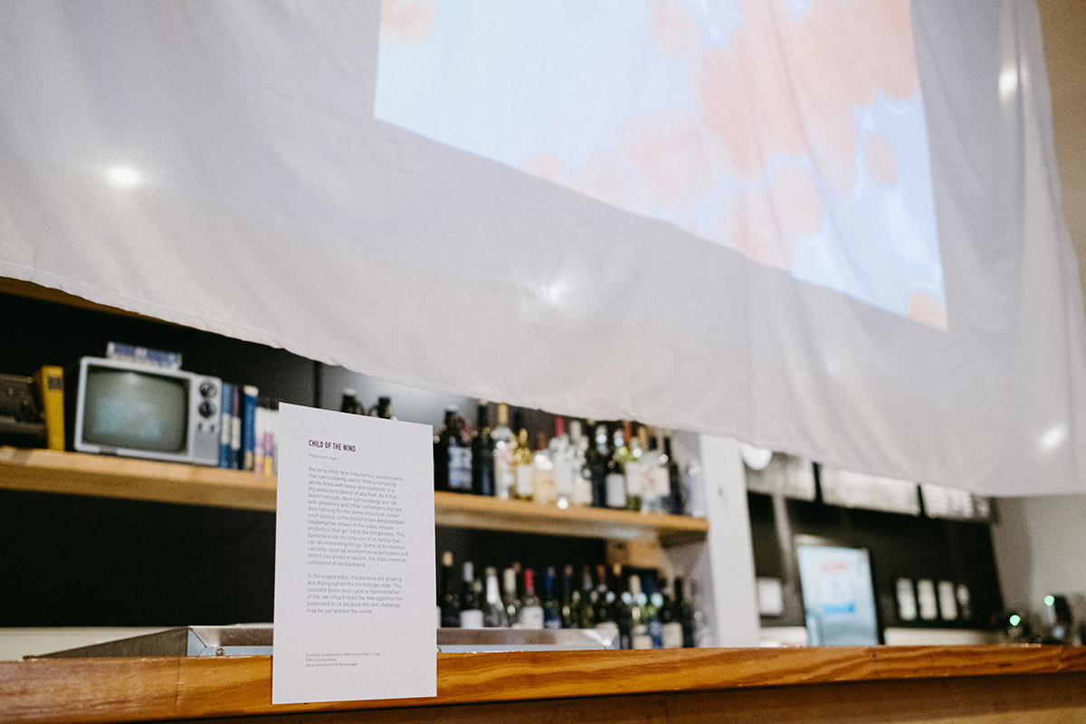
Child of the Wind
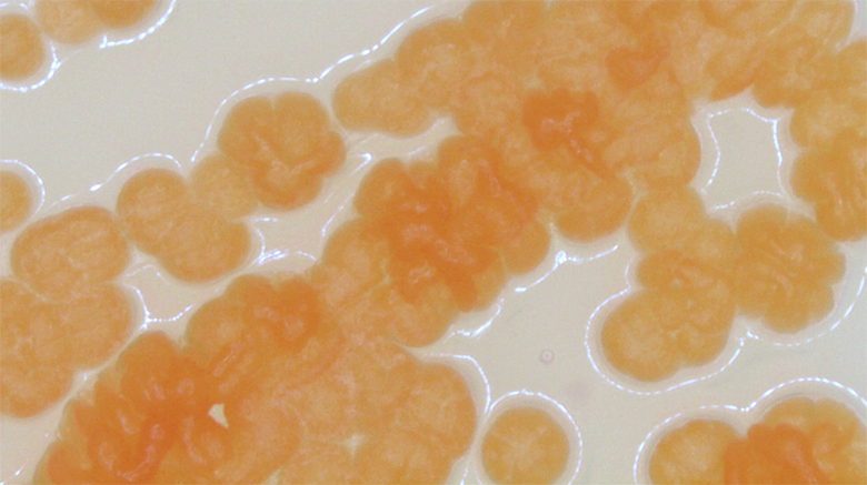
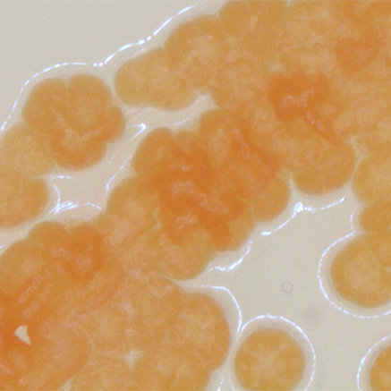
Bacteria often face treacherous environments that can suddenly switch from a nurturing abode filled with water and nutrients to a dry wasteland devoid of any food. As if that wasn’t enough, their surroundings are rife with predators and other contenders that are also fighting for the same resources. Under such duress, some bacteria like Amycolatopsis tolypomycina, shown in the video, release antibiotics that get rid of the competition. This bacteria is not the only one in its family that can do interesting things. Some of its relatives can help clean up environmental pollutants and others can produce vanillin, the main chemical compound of vanilla beans.
In the looped video, the bacteria are growing and drying out on the microscope slide. This constant boom-bust cycle is representative of life: we should seize the new opportunities presented to us because the next challenge may be just around the corner.
Scientist Collaborators
Jim Dalling and Mayandi Sivaguru
IGB Core Facilities
Instrument
NSI X5000 High Resolution CT
Funding Agency
IGB Core Facilities
Original Imaging

Special Thanks
Kleinmuntz Center for Genomics in Business and Society; Nelson family and BodyWork Associates; Matt Cho and Cafeteria and Company
Image Rights
Images not for public use without permission from the Carl R. Woese Institute for Genomic Biology
Scientist Collaborators
Jim Dalling and Mayandi Sivaguru
IGB Core Facilities
Instrument
NSI X5000 High Resolution CT
Funding Agency
IGB Core Facilities
Original Imaging

Special Thanks
Kleinmuntz Center for Genomics in Business and Society; Nelson family and BodyWork Associates; Matt Cho and Cafeteria and Company
Image Rights
Images not for public use without permission from the Carl R. Woese Institute for Genomic Biology
Scientist Collaborators
Jim Dalling and Mayandi Sivaguru
IGB Core Facilities
Instrument
NSI X5000 High Resolution CT
Funding Agency
IGB Core Facilities
Original Imaging

Special Thanks
Kleinmuntz Center for Genomics in Business and Society; Nelson family and BodyWork Associates; Matt Cho and Cafeteria and Company
Image Rights
Images not for public use without permission from the Carl R. Woese Institute for Genomic Biology
Scientist Collaborators
Jim Dalling and Mayandi Sivaguru
IGB Core Facilities
Instrument
NSI X5000 High Resolution CT
Funding Agency
IGB Core Facilities
Original Imaging

Special Thanks
Kleinmuntz Center for Genomics in Business and Society; Nelson family and BodyWork Associates; Matt Cho and Cafeteria and Company
Image Rights
Images not for public use without permission from the Carl R. Woese Institute for Genomic Biology
Scientist Collaborators
Jim Dalling and Mayandi Sivaguru
IGB Core Facilities
Instrument
NSI X5000 High Resolution CT
Funding Agency
IGB Core Facilities
Original Imaging

Special Thanks
Kleinmuntz Center for Genomics in Business and Society; Nelson family and BodyWork Associates; Matt Cho and Cafeteria and Company
Image Rights
Images not for public use without permission from the Carl R. Woese Institute for Genomic Biology
Scientist Collaborator
Kazi Fazle Rabbi
Nenad Miljkovic Laboratory Group
Instrument
X5000 NSI High Resolution CT and rendered in 3D using Imaris
Funding Agency
IGB Core Facilities
Original Imaging
Special Thanks
Kleinmuntz Center for Genomics in Business and Society; Nelson family and BodyWork Associates; Matt Cho and Cafeteria and Company
Image Rights
Images not for public use without permission from the Carl R. Woese Institute for Genomic Biology
Scientist Collaborator
Joanne Vanessa Hwang
Hyunjoon Kong Laboratory Group
Instrument
X5000 NSI High Resolution CT and rendered in 3D using Imaris 3D
Original Imaging
Special Thanks
Kleinmuntz Center for Genomics in Business and Society; Nelson family and BodyWork Associates; Matt Cho and Cafeteria and Company
Image Rights
Images not for public use without permission from the Carl R. Woese Institute for Genomic Biology
Scientist Collaborators
Charul Chadha and Mayandi Sivaguru
Iwona Jasiuk Laboratory Group
Instrument
NSI X-5000 3D CT system and rendered using the NSI efX software
Original Imaging
Special Thanks
Kleinmuntz Center for Genomics in Business and Society; Nelson family and BodyWork Associates; Matt Cho and Cafeteria and Company
Image Rights
Images not for public use without permission from the Carl R. Woese Institute for Genomic Biology
Scientist Collaborator
Gelson J. Pagan-Diaz
Holonyak Micro & Nanotechnology Laboratory
Instrument
Zeiss LSM 710 Confocal Microscope
Original Imaging

Special Thanks
Kleinmuntz Center for Genomics in Business and Society; Nelson family and BodyWork Associates; Matt Cho and Cafeteria and Company
Image Rights
Images not for public use without permission from the Carl R. Woese Institute for Genomic Biology
Scientist Collaborator
Gelson J. Pagan-Diaz
Holonyak Micro & Nanotechnology Laboratory
Instrument
Zeiss LSM 710 Confocal Microscope
Original Imaging
Special Thanks
Kleinmuntz Center for Genomics in Business and Society; Nelson family and BodyWork Associates; Matt Cho and Cafeteria and Company
Image Rights
Images not for public use without permission from the Carl R. Woese Institute for Genomic Biology
Scientist Collaborator
Grace Tan
Andrew Leakey Laboratory Group
Instrument
MarSurf CM explorer
Funding Agency
US Department of Energy
Original Imaging

Special Thanks
Kleinmuntz Center for Genomics in Business and Society; Nelson family and BodyWork Associates; Matt Cho and Cafeteria and Company
Image Rights
Images not for public use without permission from the Carl R. Woese Institute for Genomic Biology
Scientist Collaborator
Grace Tan
Andrew Leakey Laboratory Group
Instrument
MarSurf CM explorer
Funding Agency
US Department of Energy
Original Imaging

Special Thanks
Kleinmuntz Center for Genomics in Business and Society; Nelson family and BodyWork Associates; Matt Cho and Cafeteria and Company
Image Rights
Images not for public use without permission from the Carl R. Woese Institute for Genomic Biology
Scientist Collaborator
Grace Tan
Andrew Leakey Laboratory Group
Instrument
MarSurf CM explorer
Funding Agency
US Department of Energy
Original Imaging
Special Thanks
Kleinmuntz Center for Genomics in Business and Society; Nelson family and BodyWork Associates; Matt Cho and Cafeteria and Company
Image Rights
Images not for public use without permission from the Carl R. Woese Institute for Genomic Biology
Scientist Collaborator
Grace Tan
Andrew Leakey Laboratory Group
Instrument
MarSurf CM explorer
Funding Agency
US Department of Energy
Original Imaging
Special Thanks
Kleinmuntz Center for Genomics in Business and Society; Nelson family and BodyWork Associates; Matt Cho and Cafeteria and Company
Image Rights
Images not for public use without permission from the Carl R. Woese Institute for Genomic Biology
Scientist Collaborators
Yee Ming Khaw and Mayandi Sivaguru
Makoto Inoue Laboratory Group
Instrument
Zeiss Axiovert 200M
Funding Agency
MI start up
Original Imaging
Special Thanks
Kleinmuntz Center for Genomics in Business and Society; Nelson family and BodyWork Associates; Matt Cho and Cafeteria and Company
Image Rights
Images not for public use without permission from the Carl R. Woese Institute for Genomic Biology
Scientist Collaborator
Chris Seward
Lisa Stubbs Labratory Group
Instrument
Multiphoton Confocal Microscope Zeiss 710 microscope
Original Imaging
Special Thanks
Kleinmuntz Center for Genomics in Business and Society; Nelson family and BodyWork Associates; Matt Cho and Cafeteria and Company
Image Rights
Images not for public use without permission from the Carl R. Woese Institute for Genomic Biology
Scientist Collaborator
Yufeng He
Megan Matthews Laboratory Group
Funding Agency
Realizing Increased Photosynthetic Efficiency (RIPE)
Original Imaging
Special Thanks
Kleinmuntz Center for Genomics in Business and Society; Nelson family and BodyWork Associates; Matt Cho and Cafeteria and Company
Image Rights
Images not for public use without permission from the Carl R. Woese Institute for Genomic Biology
Scientist Collaborator
Chris Seward
Lisa Stubbs Labratory
Instrument
Multiphoton Confocal Microscope Zeiss 710 microscope
Original Imaging
Special Thanks
Kleinmuntz Center for Genomics in Business and Society; Nelson family and BodyWork Associates; Matt Cho and Cafeteria and Company
Image Rights
Images not for public use without permission from the Carl R. Woese Institute for Genomic Biology
Scientist Collaborators
Max Simon and Glenn Fried
IGB Core Facilities
Instrument
Zeiss Axiozoom V16 Microscope
Original Imaging
Special Thanks
Kleinmuntz Center for Genomics in Business and Society; Nelson family and BodyWork Associates; Matt Cho and Cafeteria and Company
Image Rights
Images not for public use without permission from the Carl R. Woese Institute for Genomic Biology
Special Thanks
Champaign businessman Doug Nelson, President of BodyWork Associates, first proposed the idea that became Art of Science, and his continued efforts to support the exhibit made its realization possible. The IGB is also grateful to James Barham of Barham Benefit Group and [co][lab] founder Matt Cho for hosting the annual exhibit.


