Art of Science 4.0
April 10, 2014 - April 10, 2014
CURATED BY
Kathryn Faith
WHEN
December 28 - December 28
WHERE

View Gallery
1
/
10
1 / x

Let it Sink In
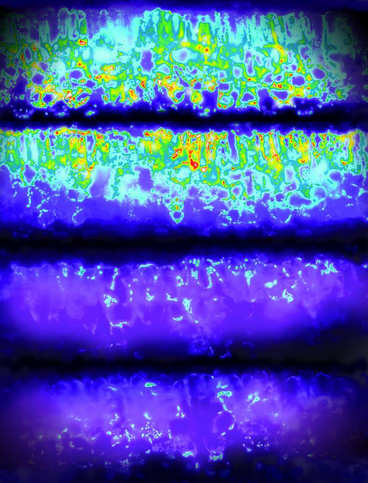
This image shows the upper and lower surfaces of a dark and a light green tobacco leaf. Fluorescent light given off by chlorophyll in cross-sections of the leaves was captured and recolored to produce the shapes seen here. Research has shown that in dark green leaves, the upper surface absorbs more light than it can use while the leaf tissue below is deprived of light. This research is testing the hypothesis that reducing chlorophyll content will result in a more efficient use of light, meaning more productive crop plants.
Power of One
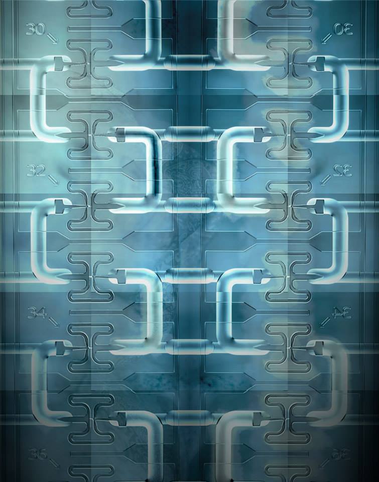
In order to measure gene expression, researchers once needed relatively large samples of tissue. Devices such as this cell capture chip made by Fluidigm now make it possible to measure gene expression in single cells. Microchannels on the chip, like the ones seen here, orchestrate the precise fluid flows needed to bring together the cells and the chemicals used to process them.
The labyrinth
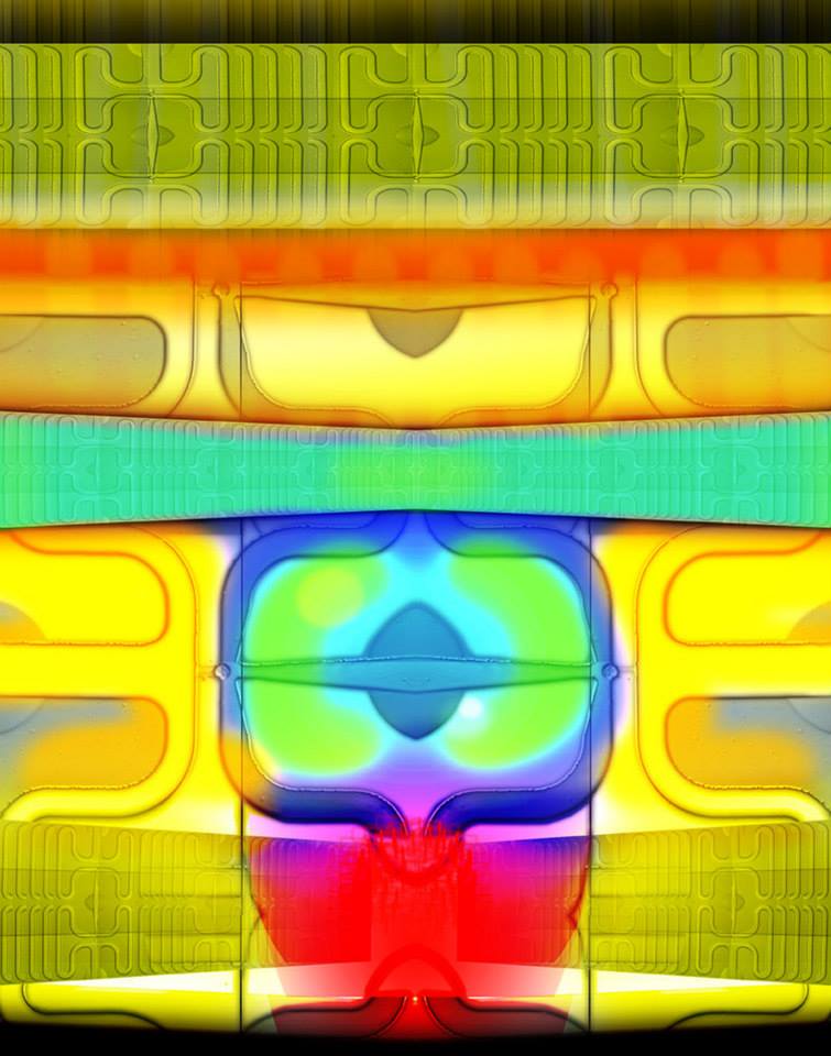
This image shows a close-up of the Fluidigm cell capture chip seen in “Power of one.” Reflected throughout the image are tiny circles, duplications of a single cell passing through one of the microchannels on the chip.
Small but mighty
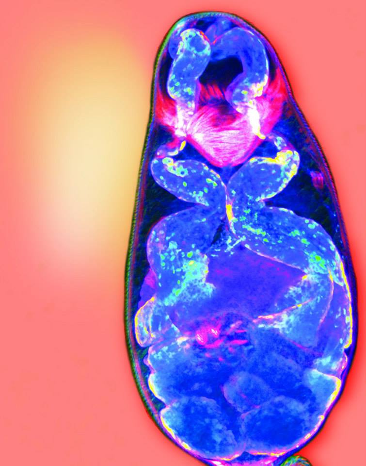
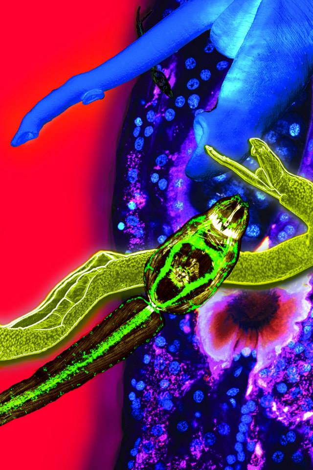
The colorful figure in this image is the larval form of a disease-causing parasitic flatworm called a schistosome. Another view of this stage of the schistosome lifecycle makes up part of the medley in the Art of Science Banner Image, “Know your enemy.” Shown in blue are glands that release chemicals that allow the worm to penetrate human skin. In red are the parasite’s muscles.
Border protection
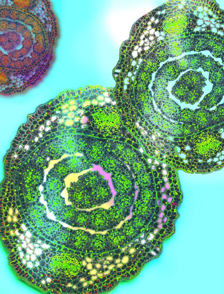
This research investigates how, where, and why certain chemicals called ‘safeners’ increase rates of herbicide detoxification in cereal crops such as grain sorghum, and how plants respond at the molecular level to safener treatment. This image illustrates a cross section of cells from a safener-treated sorghum seedling. The researchers found that safener triggers a massive expression of glutathione S-transferase (GST) proteins, which rapidly break down herbicides, mainly in the outermost cell layers.
Straining Sinew
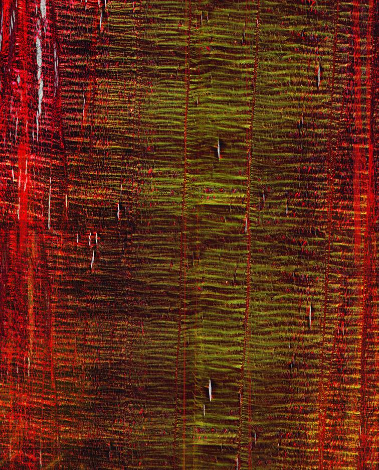
Athletic activities are hard on tendons, the connective tissue that connects muscle to bone, in people and animals alike. Tendons are made of a protein called collagen. To better understand tendon injuries, researchers injected a horse tendon with a chemical that degrades collagen. They then used images like this one, which shows the structure of the collagen fibers within the tendon, to discover how the structure is affected by damage.
Infiltration
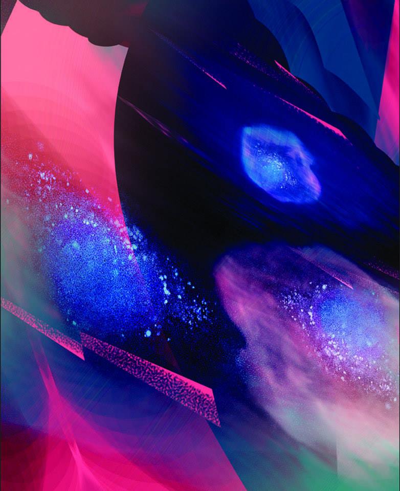
The image demonstrates that a spiral polypeptide developed by the Cheng research group, a synthetic protein, efficiently delivers DNA segments into cancer cells. This new material can potentially be used in clinical gene therapy to improve the efficiency of gene delivery, as well as to lower toxicity in healthy tissues. These are important prerequisites for the polypeptide’s future use in cancer treatment.
With a light touch
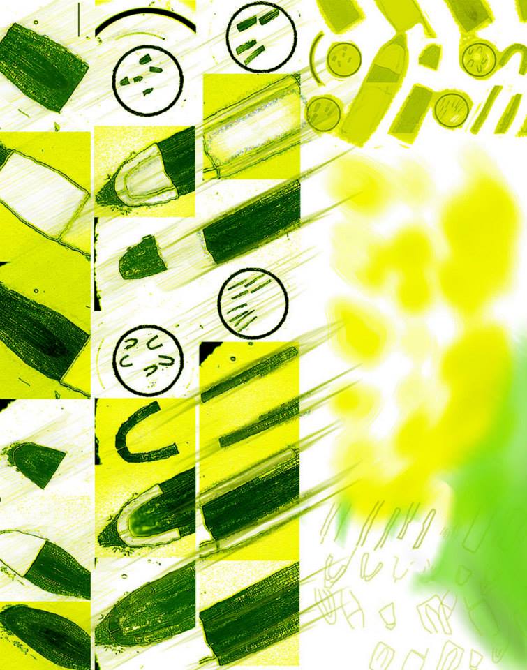
Aluminum toxicity currently affects over 40% of the land used for crops worldwide. This image shows root tips of sorghum plants treated with aluminum. Researchers use lasers to dissect out specific types of cells and tissues in treated plants in order to study the plant’s response to toxicity. In this image are several views of the dissected tissue.
Turned on its axis
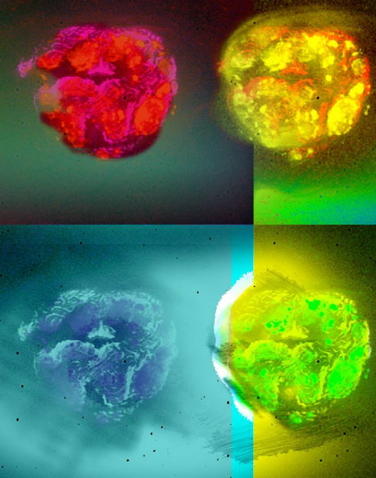
Syntaxin, a protein that plays a key role in chemical release in many types of cells, is found in the cell membrane of sperm. The head of each sperm contains an acrosome vesicle, a structure containing chemicals that help the sperm penetrate the egg. Syntaxin helps promote release of these chemicals. The research portrayed here found that syntaxin is distributed in a punctate pattern in the sperm head. Study of acrosome vesicle release will help in improving techniques used for artificial reproduction and reducing male-factor infertility.
Fire at will
Image provided by Aaron Johnson, Aaron Johnson Lab
Research funded by the University of Illinois
This repeated image shows a neuromuscular junction, the interface between a neuron and a muscle cell, located on a muscle from the larynx of a rat. With age, these laryngeal muscles become slower and weaken. By examining the laryngeal muscles in aging rats, researchers are able to investigate the effects of increased and decreased voice use on laryngeal neuromuscular mechanisms. This will help reveal if and how voice and swallowing functions can be preserved in older adults through behavioral interventions.
Web of thought
Image provided by Maminirina Randrianandrasana, May Berenbaum Lab
Research funded by the Lindbergh Foundation and the Tyler Prize for Environmental Achievement
Special thanks to Catherine Lee Wallace at the Microscopy Suite in the Imaging Technology Group at the Beckman Institute
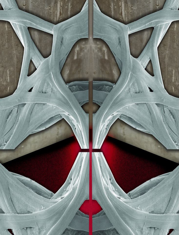
The strands running across this image are silk fibers secreted by a Madagascar wild silkworm. The structures of the fibers, observed at a microlevel scale invisible to the naked eyes, forms a mesh with crossover points. Silk is used in many enterprises, including production of cosmetics and biomedicine. Studying the properties of this particular silk is important; silk production offers an alternative income to the local people living in the border of the largest remaining forests in Madagascar, and an incentive for conserving local biodiversity.
Sweetness alight
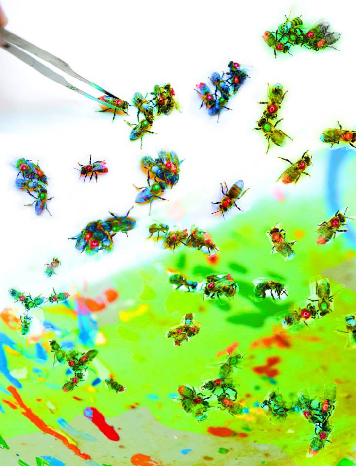
Researchers doing behavioral experiments with honey bees sometimes use paint or enamel to give individual bees distinguishing marks. The elaborate social structure and impressive learning and navigation abilities of bees make them an ideal model for behavioral and neurobiological research. Since the sequencing of the honey bee genome, published in 2006, bees have been used increasingly for research into the molecular basis for social interaction and other complex behaviors.
Living rock
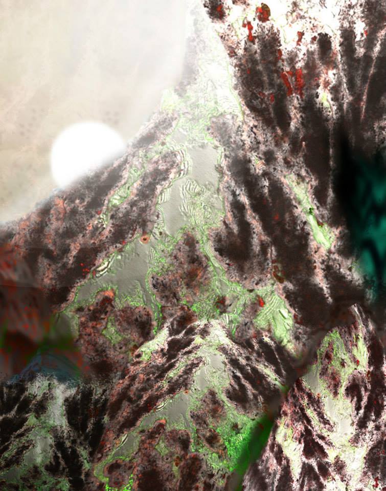
Only recently have ancient carbonate deposits that formed within hot springs (called ‘travertine’) been discovered to house globally significant volumes of subsurface oil. The successful exploration and production of these hydrocarbon reservoirs depends directly on the ability to understand and predict pore networks and permeability. Images like this, of thin travertine sections, suggest that cells line micropore walls and that the pores evolved as the result of the interplay between microbiological, physical and chemical processes.
Zeiss ELYRA superresolution microscope
Cellular Silhouettes
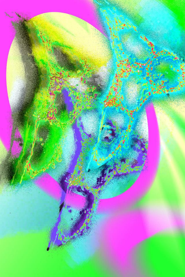
All eukaryotic cells contain highly evolved structures called mitochondria, which coordinate energy production and distribution based on the availability of calories, oxygen and the demands for cellular maintenance. In this image, a genetically encoded fluorescent protein was used to detect mitochondria. Bioenergetic reprogramming occurs in many cancer cells; genetically encoded sensors have revolutionized the study of this type of phenomenon, since they offer a convenient tool to measure bioenergetic changes in live cells.
A cell changes its spots
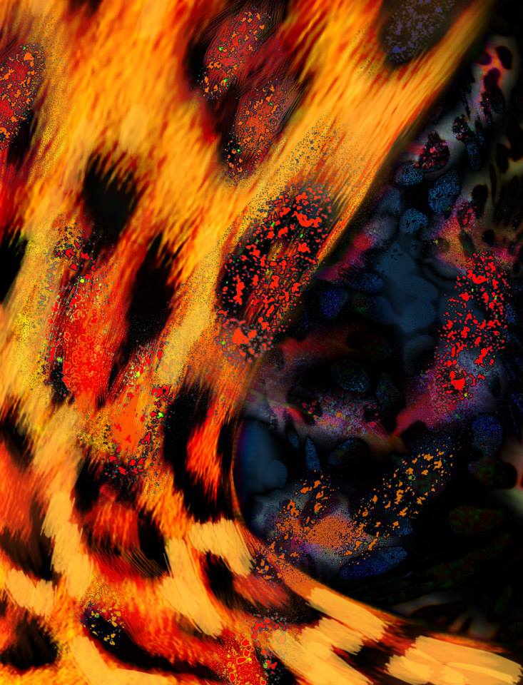
The best-known role of RNA is that of a messenger within the cell, carrying instructions for how to build proteins. However, RNA plays many other important roles, some of which are still being discovered. The bright spots that form the basis for this image were produced by labeling a long non-coding RNA (a recently described class of RNA) with a fluorescent dye. Imaging long non-coding RNAs will shed light on possible correlations between their organization in the nucleus and their potential function in coordinating gene expression.
Face of history
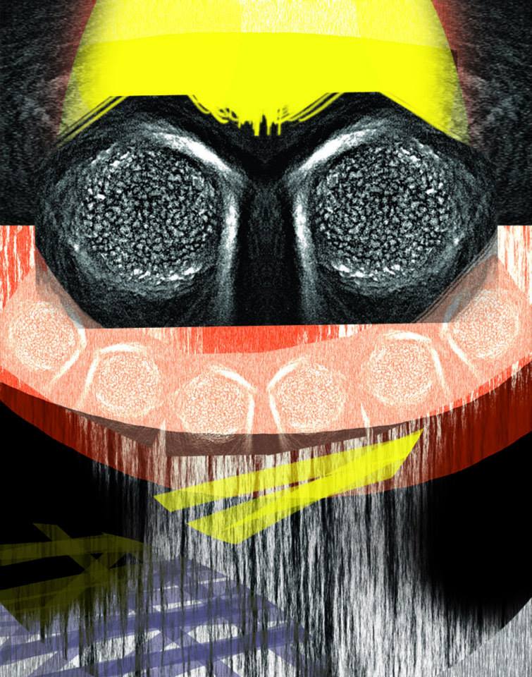
The gray texture seen here was created from the image of a single pollen grain of Themeda avenacea, oat kangaroo grass. High-power microscopy revealed surface texture features of the grass pollen too small to be seen with conventional methods; these features can be used to distinguish the species. Scientists can identify the presence of different plants from their pollen grains due to the innate morphological differences in their shape and surface texture. This practice allows scientists to reconstruct knowledge of past vegetation from modern and fossil pollen records.
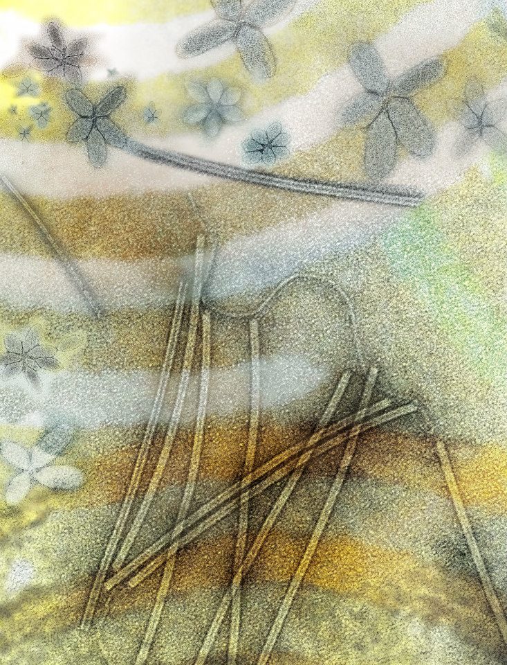
Springs eternal
These star-shaped patterns are composed of viruses that infect Sulfolobus islandicus, a species of archaea that lives in volcanic springs. The study of viruses that infect organisms of the third domain of life, the Archaea, has revealed novel and unusual filamentous and spindle-shaped viruses. The study of these viruses and how they interact with their archaeal hosts can provide insights into how early life on earth evolved.

Image provided by Becky Slattery, Don Ort Lab
Research funded by the United States Department of Agriculture
This image shows the upper and lower surfaces of a dark and a light green tobacco leaf. Fluorescent light given off by chlorophyll in cross-sections of the leaves was captured and recolored to produce the shapes seen here. Research has shown that in dark green leaves, the upper surface absorbs more light than it can use while the leaf tissue below is deprived of light. This research is testing the hypothesis that reducing chlorophyll content will result in a more efficient use of light, meaning more productive crop plants.
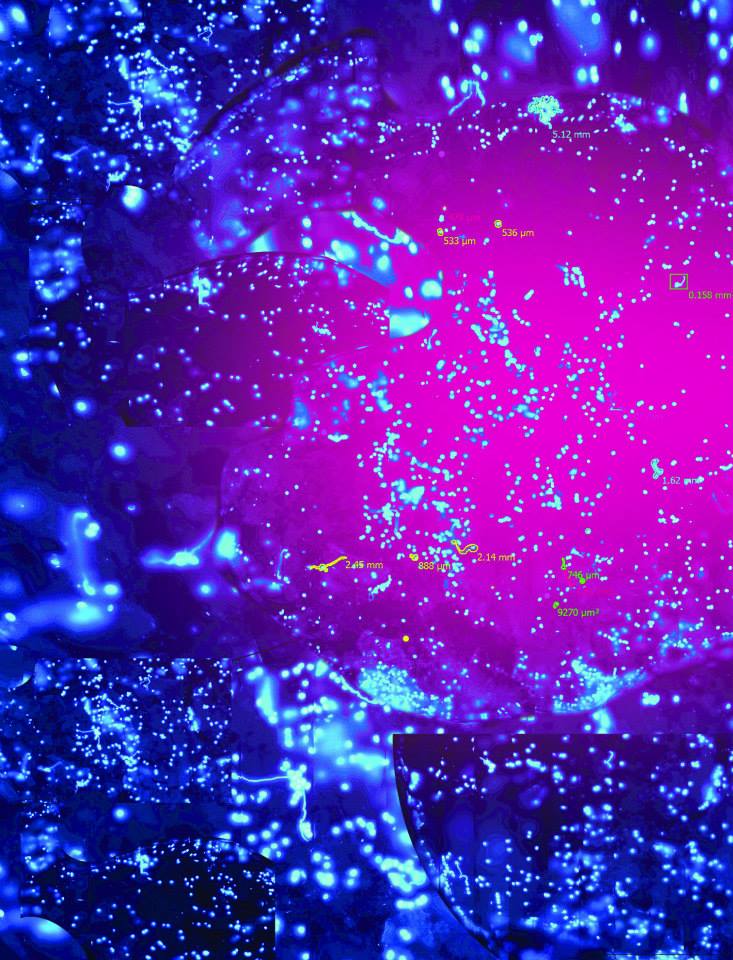
Girl Power
This image, of a germinating grain of maize pollen, was captured by a team of middle school girls participating in the Institute for Genomic Biology Pollen Power! summer camp. The camp provides an opportunity for girls to study plant responses to climate change in the distant past and the coming century. Campers watched maize pollen germinate in real-time, and learned to use Core Facilities equipment to capture and display images like this one.
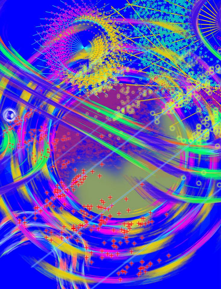
Echoes of soundness
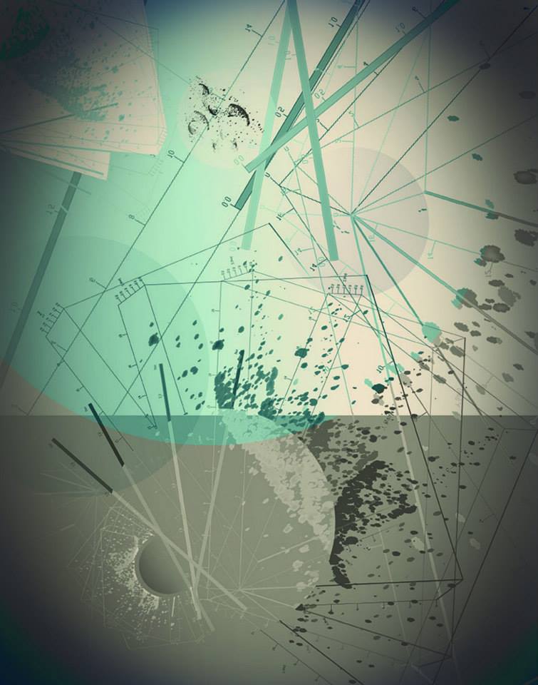
The research upon which this image is based measures health status using spatio-temporal motion by mobile devices, to draw conclusions about population health. With these data, healthy subjects and patients with chronic disease can be clearly distinguished. There are currently no reliable methods to monitor health status in everyday life; this analysis software transforms cheap phones into medical devices for viable healthcare.
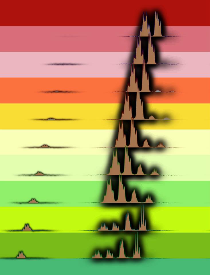
Moving picture
This series of plots represents the change in structure over time of an enzyme interacting with its substrate. Enzymes are proteins that catalyze chemical processes by interacting with the reactants. Researchers involved in drug design need information about reaction mechanisms, and how enzymes support them. Examining the changing structure of the enzyme-reactant interaction over the course of the reaction reveals important information about these mechanisms.
Know your enemy
Humans have been hosting parasitic flatworms called schistosomes for more than 5,000 years. Today these parasites continue to cause disease. This image shows several stages of the schistosome life cycle, superimposed: the egg production organs of a female (blue and purple), a larva (green and brown), and the bodies (green) and heads (blue) of adults. Understanding the life cycle could help researchers find better ways to prevent the disease the parasites cause.

The Underwater World of Curaçao
This video shows the complex and diverse ecosystem that is created by corals. Study species Montastraea faveolata was surveyed on the Caribbean island of Curaçao and appears as the abundant coral in this video. Coral reef ecosystems are critically important reservoirs of biodiversity in various marine environments, yet they are also one of the most threatened marine ecosystems on the planet. The goal of this research is to create a quantitative and qualitative baseline for what is a “healthy” coral. This baseline is vitally important as a comparative standard for determining how environmental factors affect coral health.
See the video at http://youtu.be/tbgDmRp8Riw
Let it Sink In
Scientist Collaborator
Becky Slattery
Don Ort Lab
Instrument
Zeiss Lightsheet Z.1 Microscope
Funding Agency
Research funded by the United States Department of Agriculture
Original Imaging

Special Thanks
The Labrynth
Scientist Collaborator
Mark Band
Functional Genomics Unit
Roy J. Carver Biotechnology Center
Instrument
Zeiss LSM 710 Confocal Microscope
Funding Agency
Research funded by the Roy J. Carver Biotechnology Center and the Office of Vice Chancellor of Research at the University of Illinois
Original Imaging


Special Thanks
Small but mighty
Scientist Collaborator
Jim Collins
Phil Newmark
Phil Newmark Lab
Instrument
Zeiss LSM 710 Confocal Microscope
Funding Agency
Research funded by the National Institute Health and the Howard Hughes Medical Institute
Original Imaging


Special Thanks
Border protection
Scientist Collaborator
Mayandi Sivaguru
Anatoli Lygin
Dean Riechers
Dean Riechers Lab
Instrument
Multiphoton Confocal Microscope Zeiss 710 with Mai Tai eHP Ti:sapphire laser
Funding Agency
Research funded by the University of Illinois Campus Research Board and the Cooperative State Research, Education, and Extension Service
Original Imaging


Special Thanks
Straining Sinew
Scientist Collaborator
Mayandi Sivaguru
Core Facilities
Instrument
Nanozoomer Slide Scanner
Funding Agency
Research funded by the American Quarter Horse Association
Original Imaging


Special Thanks
Infiltration
Scientist Collaborator
Yanrong Lu
Qian Yin
Nan Zheng
Jianjun Cheng Lab
Instrument
Zeiss Elyra S1SIM
Funding Agency
Research funded by the National Institutes of Health
Original Imaging


Special Thanks
With a light touch
Scientist Collaborator
Mayandi Sivaguru
Core Facilities
Instrument
Veritas Laser Capture Microdissection (LCM) and Laser Cutting System
Funding Agency
Research funded by the Unites States Department of Agricultural
Original Imaging


Special Thanks
Turned on its axis
Scientist Collaborator
Momal Sharif
David Miller Lab
Instrument
SR-SIM Four laser structured illumination system; Imaris 3D visualization package
Funding Agency
Research funded by the COMSATS Institute of Information and Technology, Islamabad, Pakistan
Original Imaging


Special Thanks
Fire at Will
Scientist Collaborator
Aaron Johnson
Aaron Johnson Lab
Instrument
SR-SIM Four laser structured Illumination system
Funding Agency
Research funded by the University of Illinois
Original Imaging


Special Thanks
Web of thought
Scientist Collaborator
Maminirina Randrianandrasana
May Berenbaum Lab
Instrument
SR-SIM Four laser structured Illumination system
Funding Agency
Environmental scanning electron microscope with a field-emission electron gun
Original Imaging


Special Thanks
Sweetness alight
Scientist Collaborator
Claudia Lutz
Charley Nye
Gene Robinson Lab
Instrument
Canon EOS 5D Mark II
Funding Agency
Research funded by the National Institutes of Health
Original Imaging


Special Thanks
Living rock
Scientist Collaborator
Eva De Boever
Mayandi Sivaguru
Bruce Fouke Lab
Instrument
Multiphoton Confocal Microscope Zeiss 710 with Mai Tai eHP Ti:sapphire laser
Funding Agency
Research funded by the TOTAL E&P Oil Company Recherche Développement
Original Imaging


Special Thanks
Cellular silhouettes
Scientist Collaborator
Vladimir Kolossov
Rex Gaskins
Rex Gaskins Lab
Instrument
Zeiss ELYRA superresolution microscope
Funding Agency
Research funded by the National Institutes of Health
Original Imaging


Special Thanks
A cell changes its spots
Scientist Collaborator
Jingyi Fei
Taekjip Ha Lab
Instrument
SR-SIM - Four laser structured illumination system
Funding Agency
Research funded by the National Science Foundation and the American Cancer Society
Original Imaging


Special Thanks
Face of history
Scientist Collaborator
Cassandra J. Wesseln
Surangi Punyasena Lab
Instrument
SR-SIM - Four laser structured illumination system
Funding Agency
Research funded by the National Science Foundation
Original Imaging


Special Thanks
Springs Eternal
Scientist Collaborator
Maria Alejandra Bautista
Rachel Whitaker Lab
Instrument
Philips CM200 transmission electron microscope
Funding Agency
Research funded by the National Science Foundation
Original Imaging


Special Thanks
Girl Power
Scientist Collaborator
Pollen Power! campers
Andrew Leakey
Core Facilities
Instrument
Nanozoomer Slide Scanner
Funding Agency
Research funded by the National Science Foundation
Original Imaging


Special Thanks
Echoes of soundness
Scientist Collaborator
Qian Cheng
Bruce Schatz Lab
Instrument
Analyze - 3D Visualization and analysis software
Funding Agency
Research funded by the United States Department of Agriculture
Original Imaging


Special Thanks
Moving picture
Scientist Collaborator
Xinshuai Zhang
John Gerlt Lab
Instrument
Agilent 600 Mhz NMR
Funding Agency
Research funded by the National Institutes of Health
Original Imaging


Special Thanks
Know your enemy
Scientist Collaborator
Jim Collins
Phil Newmark
Phil Newmark Lab
Instrument
Various instrumentation
Funding Agency
Research funded by the National Institutes of Health
and the Howard Hughes Medical Institute
Original Imaging


Special Thanks
The Underwater World of Curaçao
Scientist Collaborator
Colleen Cook
Bruce Fouke Lab
Instrument
GoPro Video Camera
Funding Agency
Research funded by the Fouke Lab Group, the Illinois Department of Geology, the Roscoe Jackson
Award for Research Travels, and the Illinois Graduate College Master’s Travel Grant
Original Imaging


Special Thanks
Special Thanks
Champaign businessman Doug Nelson, President of BodyWork Associates, first proposed the idea that became Art of Science, and his continued efforts to support the exhibit made its realization possible. The IGB is also grateful to James Barham of Barham Benefit Group and [co][lab] founder Matt Cho for hosting the annual exhibit.


