CURATED BY
Kathryn Faith
WHEN
December 16 - December 16
WHERE

View Gallery
1
/
10

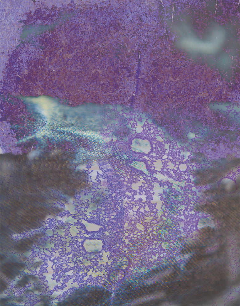
The awareness that what we eat influences our well-being predates modern science, yet we are still discovering new connections between diet and health. This image shows mouse lung tissue; the normal cells near the bottom, sampled from a healthy mouse, contrast with the cancerous cells at the top, sampled from a mouse whose diet included oil previously heated to deep-frying temperatures. Research efforts like this one help to clarify the hidden health risks and benefits of different foods.

In order to better understand and diagnose disease, bioengineers are developing living cellular machines: laboratory devices in which inorganic and organic parts are integrated. To do this, they need a way to encourage the development and interaction of different cell types. The bright reds and greens in this image come from neurons and support cells, demonstrating that researchers can successfully induce the development of specific desired cell types in the laboratory.

Fluorescent light is often used to create biological images, revealing the composition and functions of living cells. However, this type of light can negatively affect cells, sometimes causing them to detach from the substrate they are growing on. To study this phenomenon, scientists used microscope particles, which appear here as tiny points of light, to track how certain types of light exposure cause a physical relaxation of forces found in living cells.

This delicate structure is a single brain cell (neuron) from a region of the brain called the hippocampus, which contributes to the formation and recall of memories. The many elaborate branches seen here are neurites; information is exchanged between neurons at synapses, where neurites meet. Images like this one enable the study of neurite development, helping us to understand what goes wrong in diseases of brain development.

Lawrence Valverde
William Wilson Laboratory
Funded by the DOE
Work with microscopes typically relies on the intensity of emitted or reflected light. A method called fluorescence lifetime imaging microscopy is different, focusing instead on how quickly emitted light disappears. The rising and falling patterns in this image are derived from the lifetimes of fluorescent light emitted by reactants and products as they swirl together inside a tiny network of channels, providing a novel view of how these molecules interact.

The awareness that what we eat influences our well-being predates modern science, yet we are still discovering new connections between diet and health. This image shows mouse lung tissue; the normal cells near the bottom, sampled from a healthy mouse, contrast with the cancerous cells at the top, sampled from a mouse whose diet included oil previously heated to deep-frying temperatures. Research efforts like this one help to clarify the hidden health risks and benefits of different foods.

Mitochondria, the cellular structures that convert food into energy, are essential for maintaining chemical balances and signaling systems. Each dot shown here represents a single mitochondrion. Energy requirements within a tissue influence the distribution of mitochondria, and this in turn influences other activities of the cell, including disease processes and cell death. Tracking the distribution of mitochondria may suggest new ways to treat a multitude of diseases, including cancer.

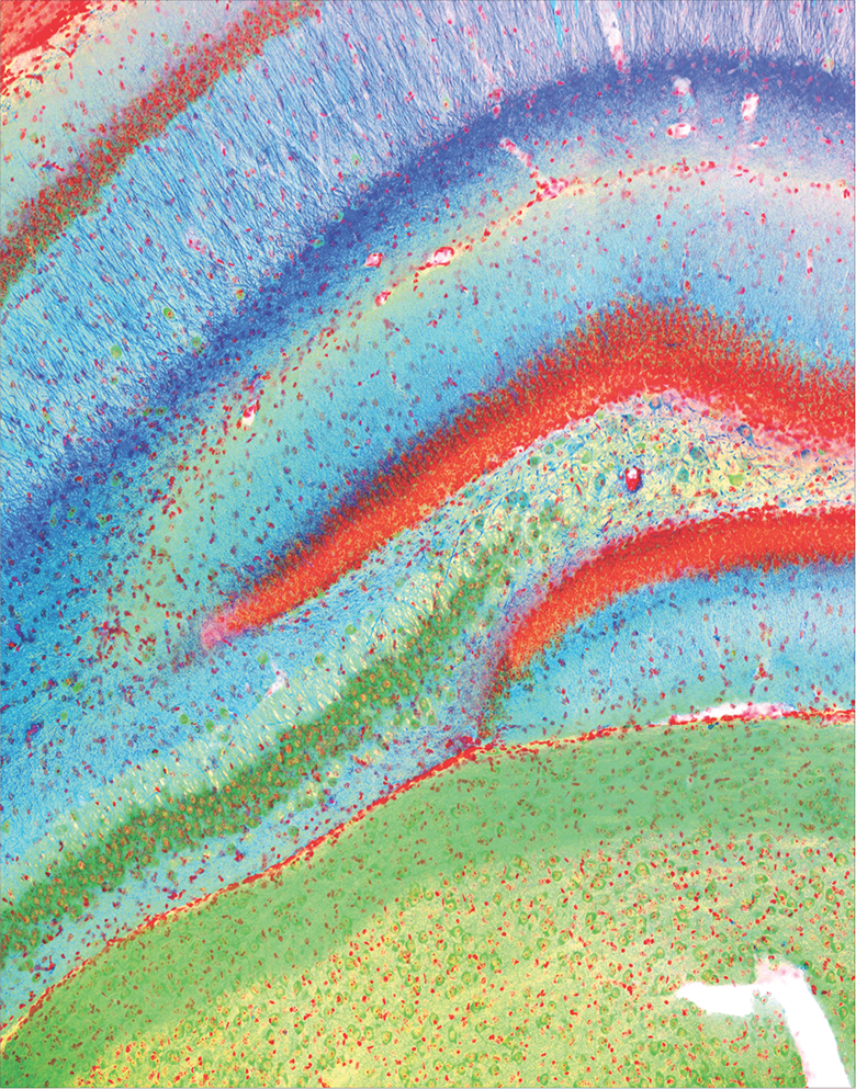
The gene AUTS2 plays a critical role in the development of the brain and has been implicated in a wide variety of human neurological disorders, including autism. Studying mice with a mutated form of this gene allows researchers to investigate how it contributes to disease pathology. The vibrant colors shown here highlight AUTS2’s activity and the important structures of the brain, allowing a closer look at when, where and how the gene functions.

Waterborne viruses cause major health problems in developing countries. Microscopic images of viruses invading and escaping cells, like the one that formed the basis for this piece, allow us to examine different portions of the virus replication cycle and identify parts of the cycle that disinfectants may be able to disrupt. This research could lead to the innovation of novel effective disinfectants and rapid methods for detection of infectious viruses in drinking water.

Scientists use sudden changes in the external environment of laboratory-grown cells like this one to probe the mechanical properties of cellular proteins and larger molecular machinery. When the concentrations of salts or other solutes are changed, the cell’s outer membrane is rapidly deformed, yet remarkably, the cell resumes function upon return to normal conditions. By studying how the complex structures inside the cell achieve this resilience, researchers hope to better understand their function in health and disease.

The sturdy shapes in this image are the cells of a sorghum plant; these cells form part of the sheath that protects the growing plant shoot. Crops are exposed to many stressful growing conditions in the field, including exposure to herbicides. The bright colors seen here mark the presence of proteins that help protect the plant from such exposures. The goal of this research is to develop new ways to protect crops from damage caused by herbicides and other stressors.

Plants capture sunlight and convert its energy into sugar, a process known as photosynthesis. One might expect that plants have evolved to capture light as efficiently as possible. Instead, many plants, including food crops, have optimized their ability to out-compete neighbors, a strategy that results in inefficient capture and use of sunlight. This image shows the arrangement of sorghum leaf cells that contribute to photosynthesis and the circulation of important molecules. By understanding and influencing this internal structure, researchers hope to develop more efficient and productive crops.

Eastern Filbert Blight is a fungal disease that attacks European hazelnut trees. The disease is challenging to manage in part because it takes months for symptoms to appear after infection. The red spine-like structures in this image are the growing filaments of the fungus invading a plant stem, shown in green with a tangle of red fibers from the plant’s vascular tissue. Documenting the fungal growth pattern will help plant biologists diagnose and control the disease.

Scattered across this image are cross-sections of a clawed frog egg. The large, dark circles mark the yolk substance that feeds the developing embryo. Clawed frogs are often used as an experimental model for studies like this one that help us understand how organisms develop and grow.

The strong lines in this image echo the sinuous shape of double-stranded DNA. Imaging such a small structure—forty thousand times narrower than a single human hair—requires a very specialized instrument. This image was created as an early test of a new atomic force microscope, which uses a minuscule and precisely controlled physical probe to construct images of structures that were previously beyond the reach of scientific instrumentation.

Genetic inheritance, childhood experience, and adult environment all play important roles in human behavior, including neurological disorders with behavioral components. The network of interconnected nodes seen here represent a statistical model that quantifies the relationships between early life stress and victimization, impulse control, and binge drinking. Elucidating the lasting effects of early life stress can inform policy, advocacy, and intervention practices in an attempt to help protect our youth from long-term problems.

Biologists can never observe first-hand the evolutionary history of Earth’s diverse lifeforms. Instead, they play detective, examining the clues past processes have left behind to trace progressive changes in form and function. In this cross-section of an opossum skull, researchers can identify the similarity between the bone structure of the reptilian jaw and a developmental stage of mammalian middle ear bones. By studying middle ear development in mammals, we can better understand how the same bones used for chewing in a common ancestor became adapted for hearing in opossums and people.

The contrasting geometric shapes in this image were formed by the boundaries between a drop of sea surface seawater from the North Sea combined with a drop of oil from deep beneath the sea floor in the North Sea. Researchers constructed an experimental test bed called the GeoBioCell to study the interactions between water, rock, microbes, and oil under high magnification. Researchers hope to develop a method to prevent oil “souring,” the contamination of oil fields by microbially-produced sulfide gas.

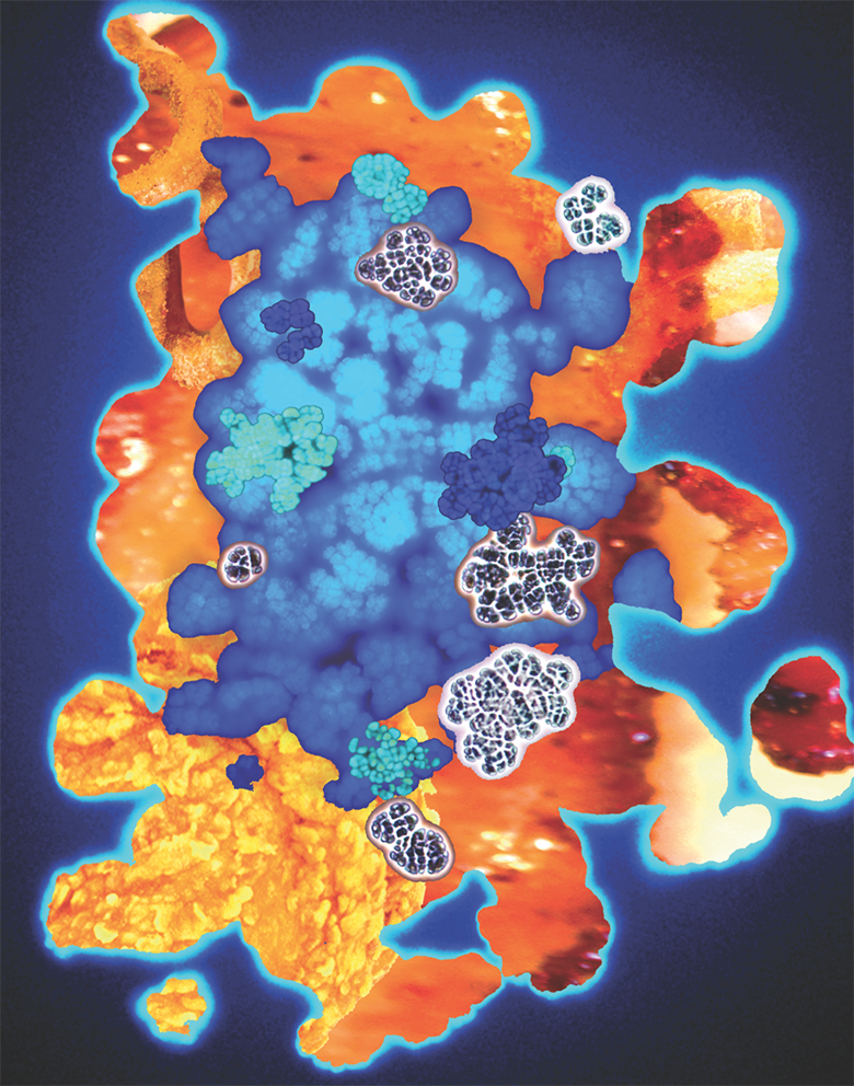
Methanosarcina is a genus of bacteria named for the shapes produced by clusters of individual cells, such as those seen in this image. These clusters resemble sarcina, the traveling packs carried by Roman soldiers; they can also resemble billowing clouds, a reminder of the contribution of these bacteria to climate change. Microbes such as these produce approximately 2 billion tons of methane each year as they decompose organic waste. Knowledge of their natural history can help us improve biofuel production while also mitigating the human contribution to climate change.

In this image, a few dark points stand out against a softer, textured background. These points represent individual copies of a single gene within human cancer cells. The researchers who created this image developed a new method that allows them to track, in 3D, the location of individual genes within cells. This tool will allow researchers to visualize fundamental biological processes and reveal how these processes are disrupted in diseases such as cancer.

The circular, branching patterns seen here are a common way to represent the evolutionary relationships between different organisms. In this research, scientists examined the relationships among species of Eukarya, the branch of life that includes animals, plants, and fungi, and Archaea, a branch of life that comprises single-celled microbes that often live in extreme environments. By comparing the DNA sequences of key genes in modern organisms, we can trace the evolution of the earliest forms of life billions of years ago.
Moirai
Scientist Collaborator
Anthony Cam
William Helferich Laboratory
Instrument
NanoZoomer Slide Scanner
Funding Agency
Funded by the NIH and the USDA
Original Imaging


Special Thanks
Image Rights
Images not for public use without permission from the Carl R. Woese Institute for Genomic Biology.
Branching Stream
Scientist Collaborator
Caroline Cvetkovic
Rashid Bashir Laboratory
Instrument
Multiphoton Confocal Microscope Zeiss 710 with Mai Tai eHP Ti:sapphire laser
Funding Agency
Funded by the NSF
Original Imaging


Special Thanks
Distress Signals
Scientist Collaborator
Samantha Knoll
Taher Saif Laboratory
Instrument
Zeiss Axiovert 200M Microscope
Funding Agency
Funded by the NSF and the Linda Su-Nan Chang Sah Doctoral Fellowship
Original Imaging


Special Thanks
Seedling of Thought
Scientist Collaborator
Rajashekar Iyer
Martha Gillette Laboratory
Instrument
Zeiss LSM 880 Airyscan
Funding Agency
Funded by the NSF and the NIH
Original Imaging


Special Thanks
Early Leaf
Scientist Collaborator
Lawrence Valverde
William Wilson Laboratory
Instrument
Multiphoton Confocal Microscope Zeiss 710 with Mai Tai eHP Ti:sapphire laser
Funding Agency
Funded by the DOE
Original Imaging


Special Thanks
Fast and Huge and Interconnected
Scientist Collaborator
Chih-Ying Chen
Lisa Stubbs Laboratory
Instrument
Zeiss LSM 880 Airyscan
Funding Agency
Funded by the NIH
Original Imaging


Special Thanks
Reservoir
Scientist Collaborator
Kelley Goncalves
Joanna Shisler Laboratory
Instrument
Zeiss LSM 880; Imaris 3D visualization package
Funding Agency
Funded by the Institute for Sustainability, Energy, and Environment
Original Imaging


Special Thanks
The Reed That Bends
Scientist Collaborator
Shahar Sukenik
Martin Gruebele Laboratory
Instrument
Zeiss Elyra S1 Super Resolution Structured Illumination Microscope
Funding Agency
Funded by the NSF
Original Imaging


Special Thanks
Temple of Demeter
Scientist Collaborator
Rong Ma, Yousoon Baek, Mayandi Sivaguru, and Dean Riechers
Dean Riechers Laboratory
Instrument
Zeiss LSM 880
Funding Agency
Funded by the USDA
Original Imaging


Special Thanks
Machinery of the Ghost
Scientist Collaborator
Aleel Grennan and Kingsley Boateng
Donald Ort Laboratory
Instrument
Zeiss Sigma VP 3View Serial Block-Face Scanning Electron Microscope
Funding Agency
Funded by the DOE
Original Imaging


Special Thanks
Mysterious Life
Scientist Collaborator
Ronald Revord, Santiago Mideros, Sivaguru Mayandi, and Frank Zhao
Sarah Lovell Laboratory
Instrument
Multiphoton Confocal Microscope Zeiss 710 with Mai Tai eHP Ti:sapphire laser
Funding Agency
Funded by the Illinois Department of Agriculture and the generosity of Melissa Meador
Original Imaging


Special Thanks
Neubauten
Scientist Collaborator
Aurora Turgeon
Jing Yang Laboratory
Instrument
Zeiss Sigma VP 3View Serial Block-Face Scanning Electron Microscope
Funding Agency
Funded by NIH
Original Imaging


Special Thanks
Cambion
Scientist Collaborator
Jaya Yodh
IGB Core Facilities
Instrument
Asylum Research Cypher ES Atomic Force Microscope
Funding Agency
Funded by NSF
Original Imaging


Special Thanks
Flight Into Self
Scientist Collaborator
Jordan Davis
Brent Roberts Laboratory
Instrument
Mplus 7.3 statistical modeling software
Funding Agency
Funded by NIH
Original Imaging


Special Thanks
Same As It Ever Was
Scientist Collaborator
Daniel Urban
Karen Sears Laboratory
Instrument
NanoZoomer Slide Scanner
Funding Agency
Funded by NSF
Original Imaging


Special Thanks
All Drains Lead to the Ocean
Scientist Collaborator
Bruce Fouke
Bruce Fouke Laboratory
Instrument
Research funded by British Petroleum and the Energy Biosciences Institute
Funding Agency
Funded by NSF
Original Imaging


Special Thanks
Marching On
Scientist Collaborator
Thomas Mand and Mary Beth Metcalf
Bill Metcalf Laboratory
Instrument
Olympus BX60 Microscope/Olympus DP72 Camera
Funding Agency
Funded by DOE
Original Imaging


Special Thanks
The Measure of Every Part
Scientist Collaborator
Ipek Tasan
Huimin Zhao Laboratory
Instrument
Zeiss Elyra S1 Super Resolution Structured Illumination Microscope
Funding Agency
Funded by NIH
Original Imaging


Special Thanks
Life’s Vintage
Scientist Collaborator
Celia Mendez and Carla Baptista
Isaac Cann Laboratory
Instrument
Analytical software
Funding Agency
Funded by NASA
Original Imaging


Special Thanks
Special Thanks
Champaign businessman Doug Nelson, President of BodyWork Associates, first proposed the idea that became Art of Science, and his continued efforts to support the exhibit made its realization possible. The IGB is also grateful to James Barham of Barham Benefit Group and [co][lab] founder Matt Cho for hosting the annual exhibit.


