
Art of Science 2.0
April 10, 2012 - April 10, 2012
Share
CURATED BY
Kathryn Faith
WHEN
March 14 - March 14
WHERE

View Gallery
1
/
20
1 / 20
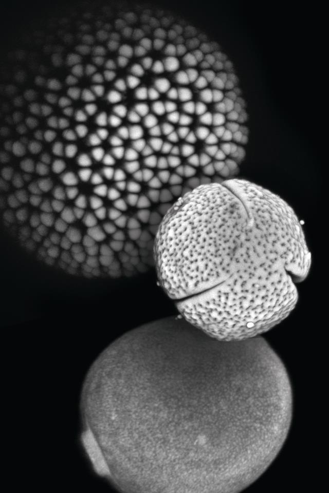
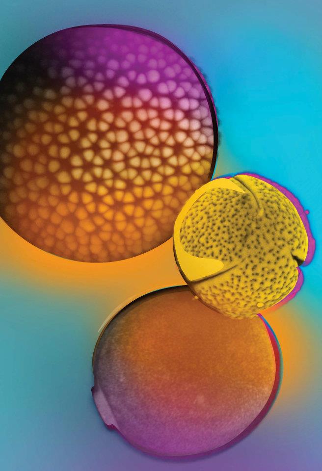
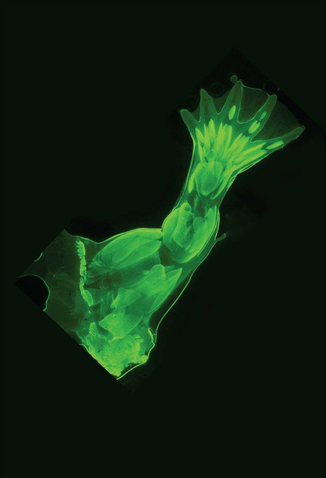
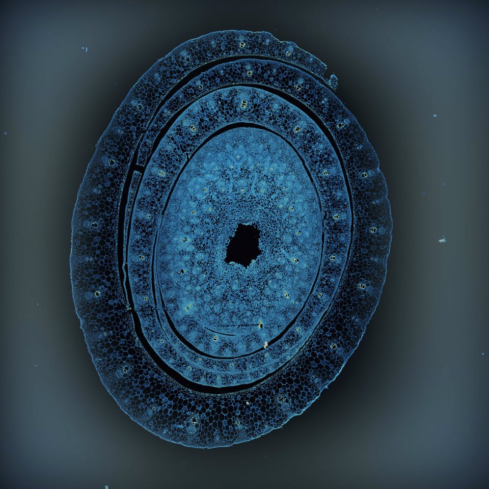
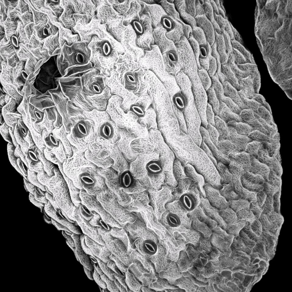
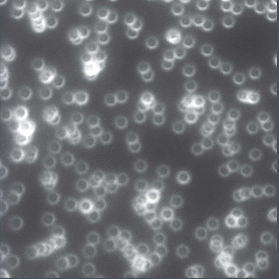
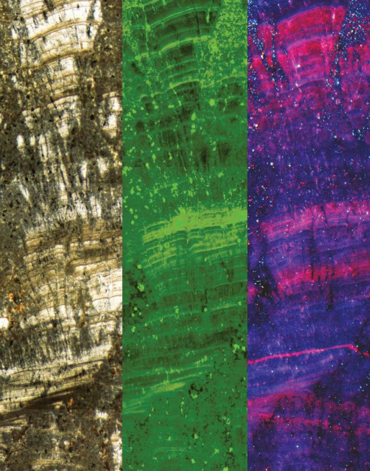
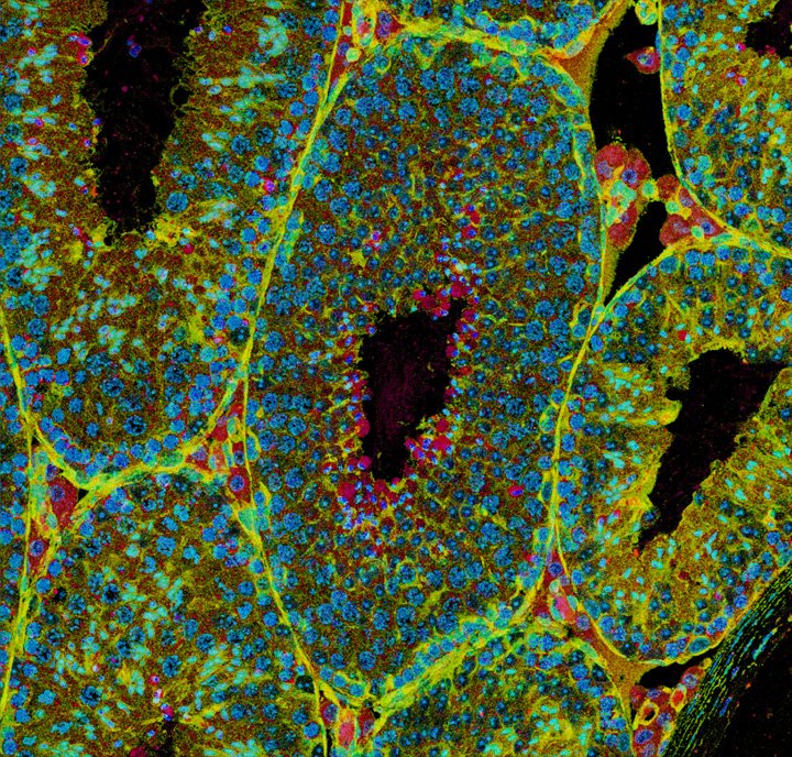
Floral Fingerprints
In this image, pollen grains from Croton hirtus (rushfoil), Mabea occidentalis (Mabea) and Agropyron repens (quackgrass) provide a glimpse of the extraordinary morphological variety of pollen. Their shapes and surface textures were revealed using cutting-edge fluorescence microscopy at the IGB. The shape of pollen grains from different plants can be so distinct that pollen identification based on structure has been used in forensic investigations, archaeological studies, and to confirm the purity of monofloral honeys.
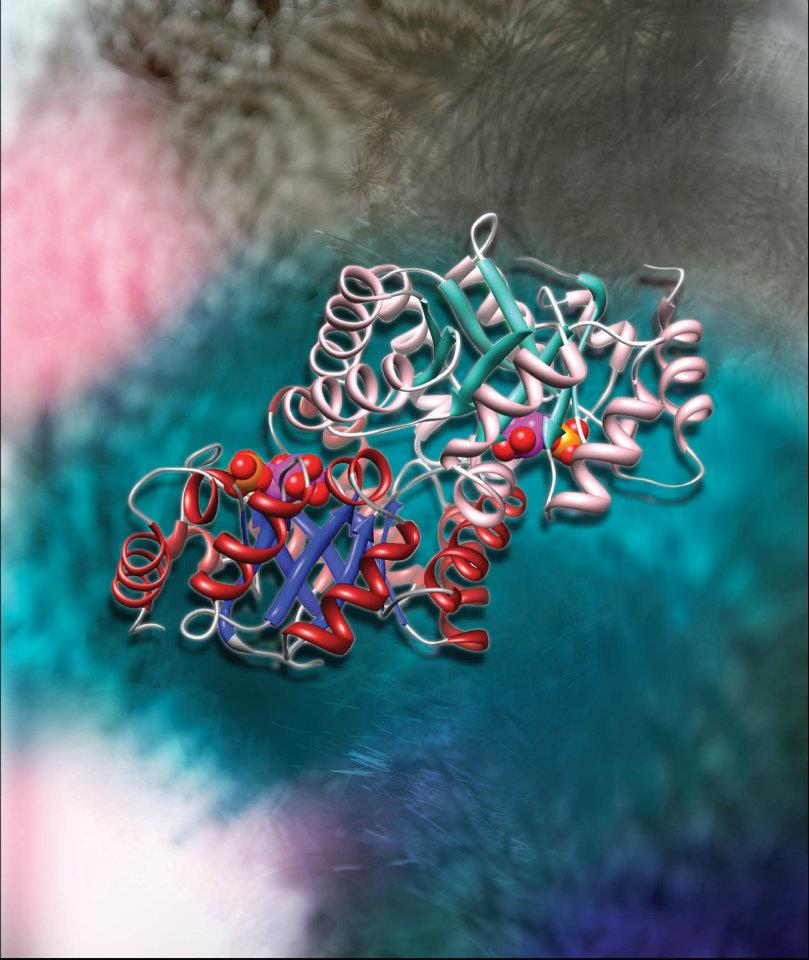
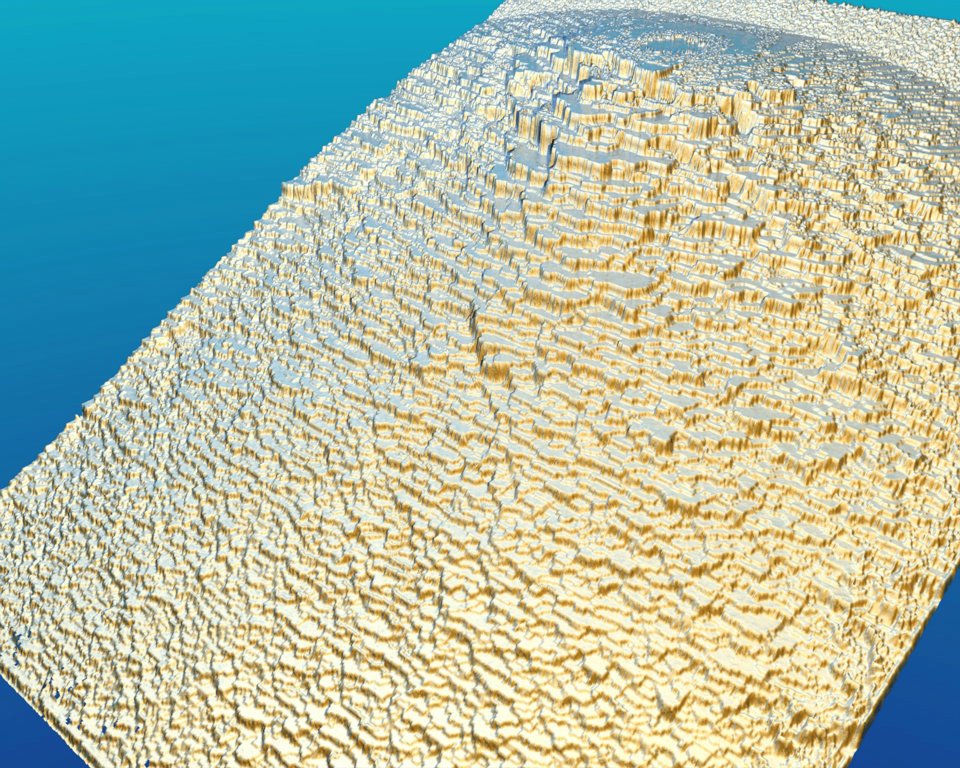
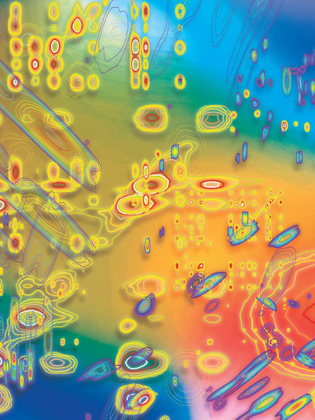
This image shows a Nuclear Magnetic Resonance (NMR) spectrum, a representation of how atoms within a molecule respond to a magnetic field. NMR spectra are very important for discovering the structure of organic compounds and proteins. They are also very useful in studying the interactions between drug molecule and target protein, information that can then be used for the structure-based drug design.
Floral Fingerprints
Scientist Collaborator
Luke Mander
Surangi Punyasena Lab
Instrument
Zeiss Apotome, Zeiss LSM 710 NLO
Funding Agency
Research funded by the National Science Foundation
Original Imaging


Image Rights
Images not for public use without permission from the Carl R. Woese Institute for Genomic Biology.
Share
Good vibrations
Scientist Collaborator
Xudong Guan
Core Facilities
Instrument
Agilent 600 MHz NMR
Funding Agency
Research funded by the Institute for Genomic Biology
Original Imaging
Image Rights
Images not for public use without permission from the Carl R. Woese Institute for Genomic Biology.
Share
Special Thanks
Champaign businessman Doug Nelson, President of BodyWork Associates, first proposed the idea that became Art of Science, and his continued efforts to support the exhibit made its realization possible. The IGB is also grateful to James Barham of Barham Benefit Group and [co][lab] founder Matt Cho for hosting the annual exhibit.