CURATED BY
Kathryn Faith
WHEN
December 2 - December 2
WHERE
View Gallery
1
/
10
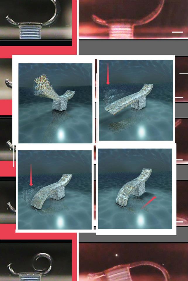
This centimeter-long bio-bot was made with a 3-D printer out of flexible polymer and living heart cells. The cells beat together, creating a contracting and releasing motion that inches the bot forward. Researchers would like to make smarter bio-bots that could assist in surgery, or find and neutralize toxins or parasites.
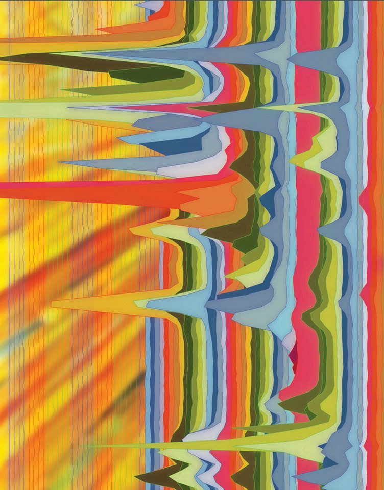
This image represents the energy released by the protons in a molecule extracted from bacteria, which is detected by sweeping the molecule with radio frequency in the presence of a magnetic field. The spectrum gives the researchers information about the connections between the atoms that make up the molecule, revealing its chemical structure.
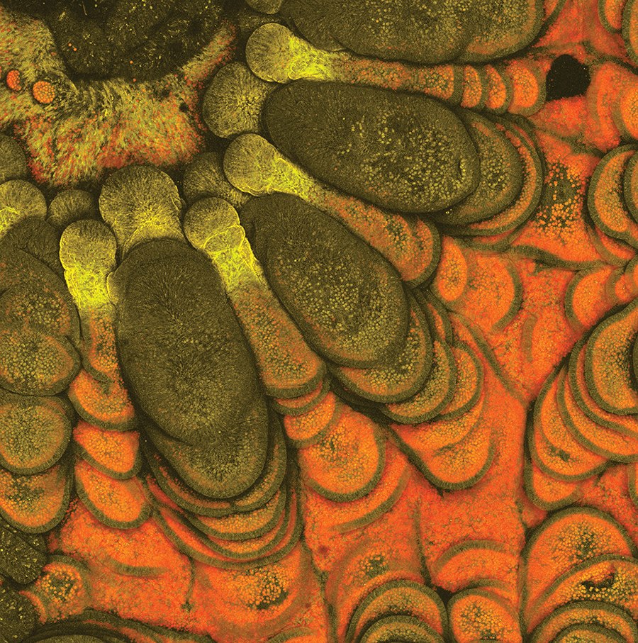
Most corals consist of many small polyps living together in a large group. This image shows cells that make up the tentacles and wall of a single polyp; part of the polyp’s mouth is visible in the upper left. Images like this one are used to understand how corals coevolved with algae to optimize the harvesting of light from seawater, and to help predict how corals will respond to global climate change.
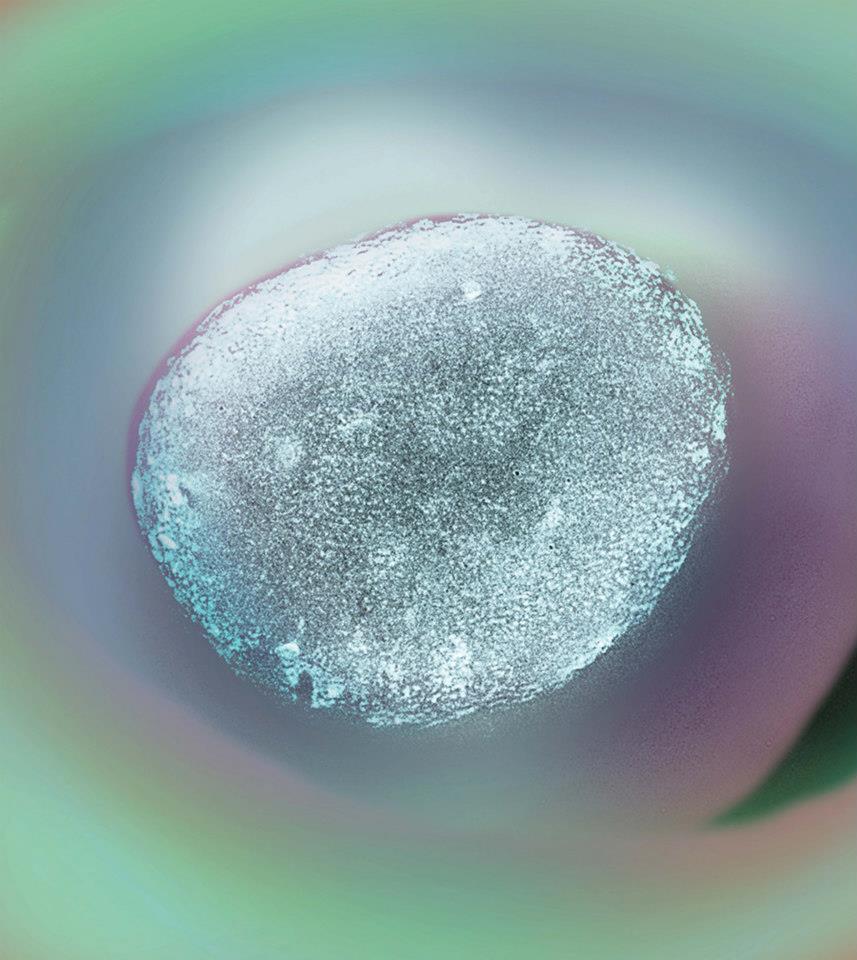
Pollen grains are an indispensable record of vegetation from the Earth’s past, and provide a unique window into the nature and timing of plant evolution. Pollen grains are also objects of considerable beauty, and their diversity of form has fascinated scientists since Malpighi and Grew first described them in the late 1600s.
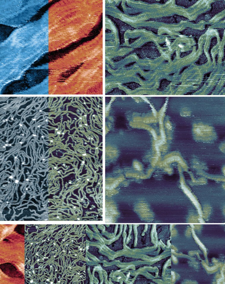
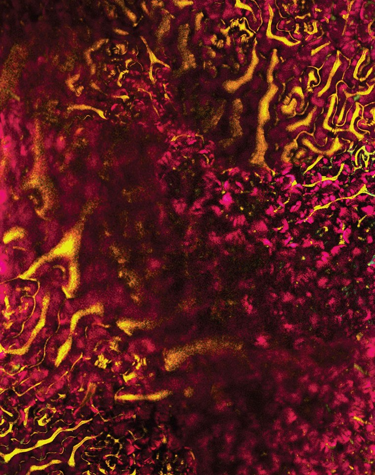
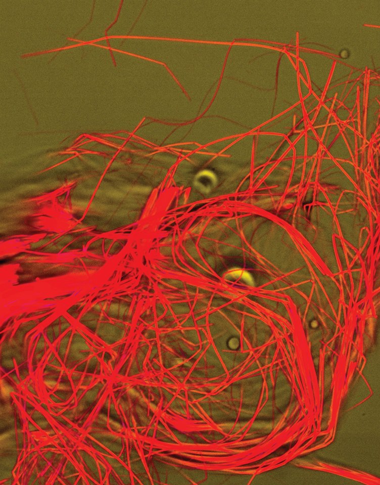
The sinuous structures shown here are made of DNA, a molecule that encodes the genetic instructions used in the development and functioning of all known living organisms. The instrument used reveals DNA’s helix structure, seen in these images as a striped pattern. These images were collected with Sophia Hohlbauch from Asylum Research.
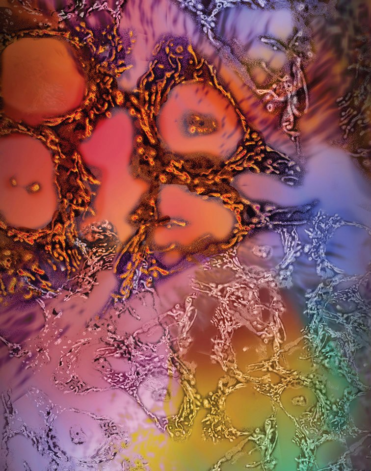
In this image of human colorectal cancer cells, the glowing points represent the chemical activity of mitochondria. Mitochondria are tiny structures that convert the energy from food into a chemical form that keeps cells running. To produce this image, researchers used a genetically encoded, chemically sensitive dye that allows them to monitor the activity of mitochondria in real time.
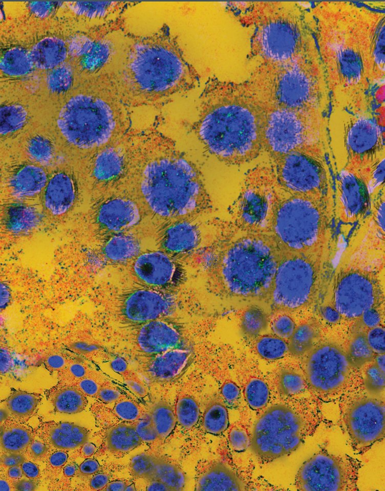
These colorful blue spots are actually sperm developing inside the testis of a male mouse. Pictures of growing sperm structure reveal how an omega-3 fatty acid, docosahexaenoic acid (DHA), contributes to sperm development. This work builds our understanding of how DHA acts in the body, leading to new insights into Alzheimer’s disease, diabetes, and the development of a male contraceptive.
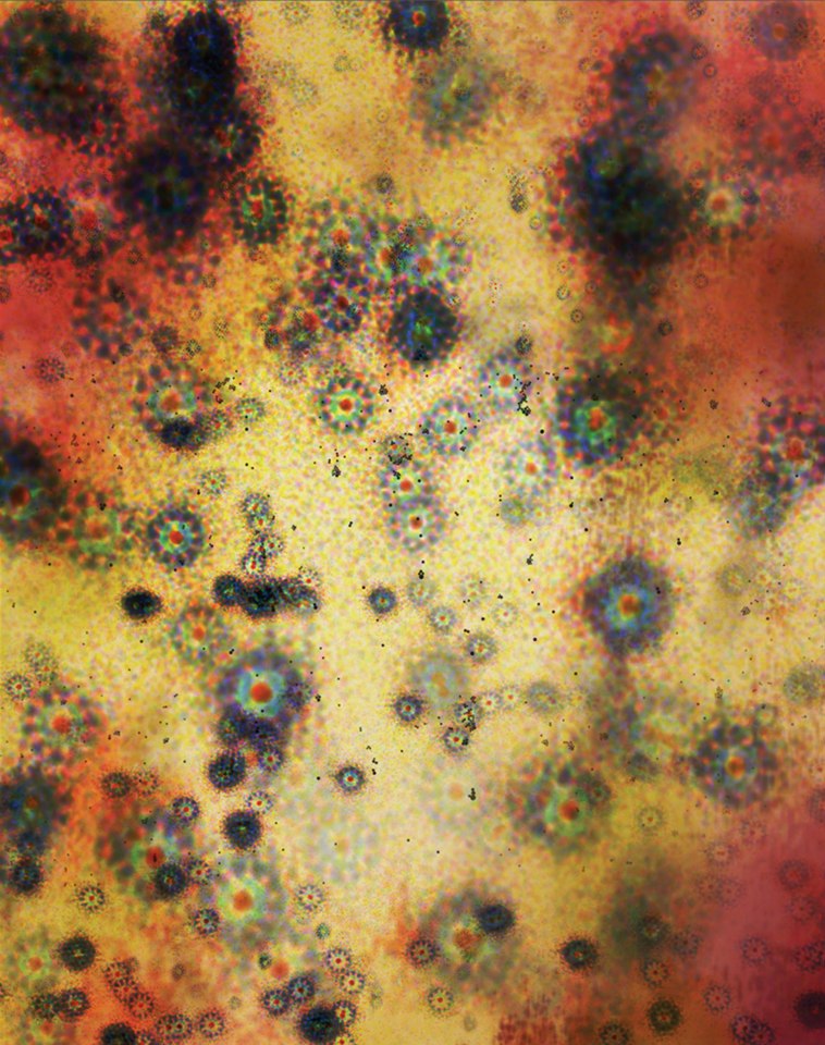
This image is a test for a research instrument. Fluorescent beads that are only 0.000017 centimeters across are used as a calibration standard to measure the limit of the optical system’s ability to produce a clear image in blue, green, orange and red wavelengths.
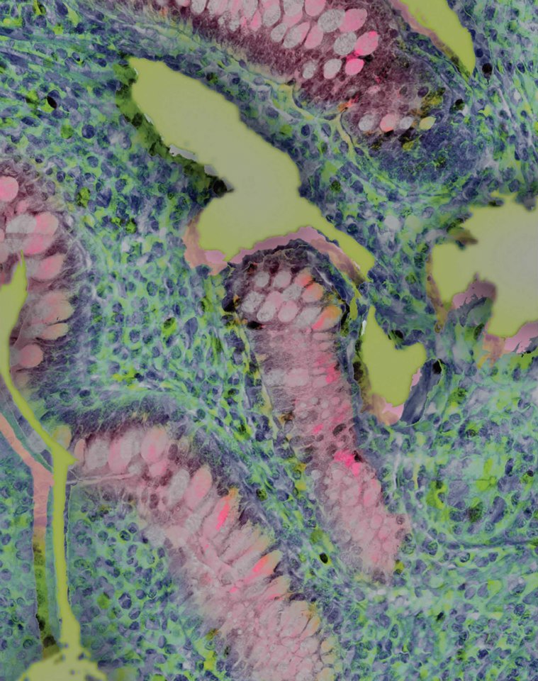
These gentle pastel colors mark dividing and maturing cells from the intestine of a young calf. Researchers are trying to identify dietary means to enhance gut health in newborn animals, in this case newborn calves, which suffer frequently from intestinal health breaks. This image provides a way to assess whether intestinal development can be facilitated by dietary stimulation.
MT1-MMP is a protein found on the membrane of metastatic cancer cells. By collecting a series of thin images and building them into the 3-dimensional images like this one, scientists observed previously unknown activation patterns of MT1-MMP. With further improvements in imaging systems, researchers aim to eventually be able to monitor in 3-D the activity of MT1-MMP as a live cancer cell travels through the body.
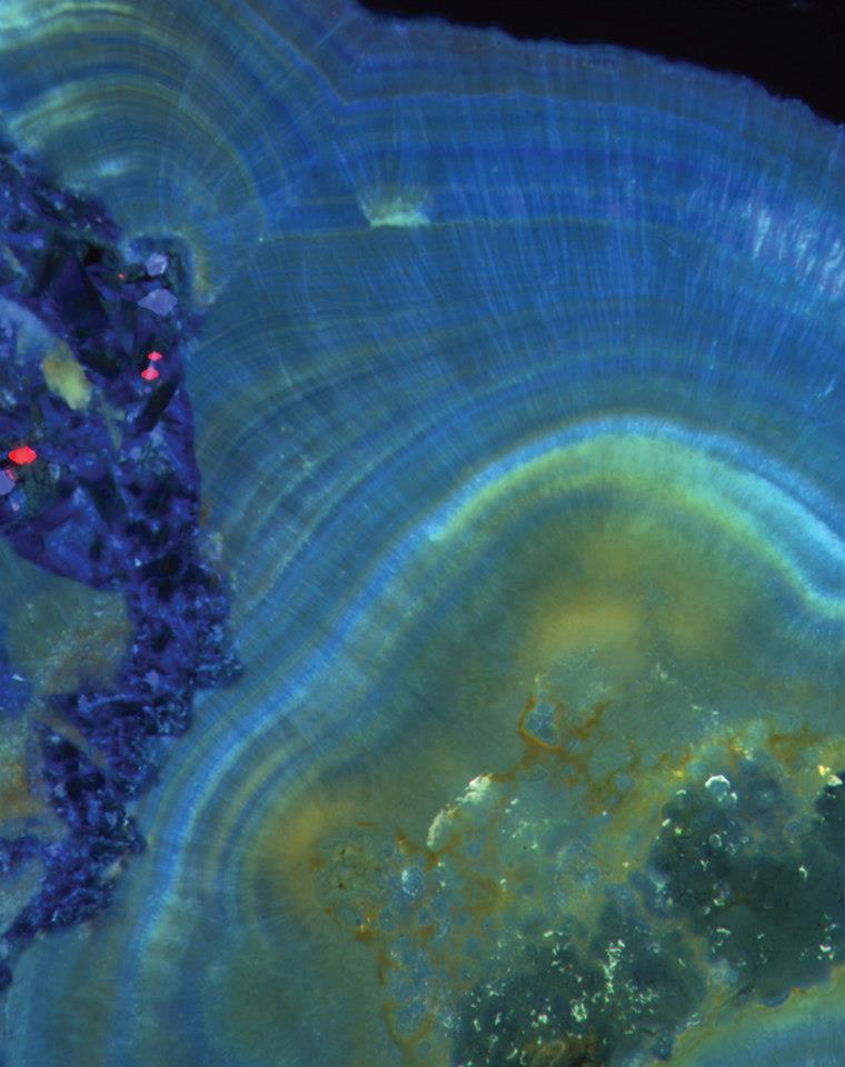
This cross-section of a human kidney stone uncovers the layers of crystal growth that were deposited during its formation. Human kidney stones have been assumed to be the product of the simple growth of crystals in naturally sterile kidneys. Images like this one suggest the opposite, that they are instead formed through complex events that include the activity of newly discovered microbes in the kidneys.
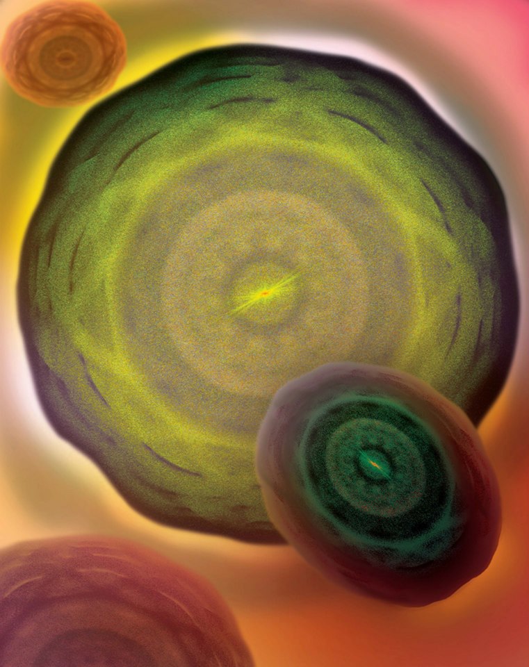
Cells are a basic unit of life because each one holds a copy of an organism’s genetic information. The images of cells shown here are not displayed in a conventional way; instead, a 3-D fast fourier transformation of the original visual information has been computed. This technique is used to show high frequency information, usually unavailable to conventional microscopy, in a visible way.
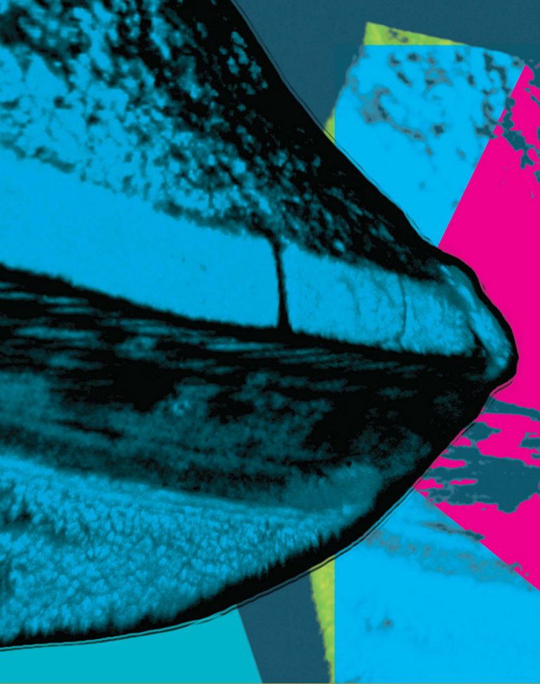
This image shows the 3-dimensional structure of the tip of a tungsten carbide micro-drill that was labeled with a fluorescent dye. The laboratory that produced this image works to create and improve micro-tools that could be used for more efficient and precise manufacturing.
This image shows an Arabidopsis leaf at different depths into the tissue. Surface views show pores that allow the leaf to “breathe.” Deeper views show a red fluorescence produced by chlorophyll. Chloroplasts closer to the surface of the leaf reach the limit of light absorption earlier than those below. By genetically reducing chlorophyll content, researchers hypothesize they may be able to create a more even distribution of light in the leaf and therefore increase photosynthetic efficiency.
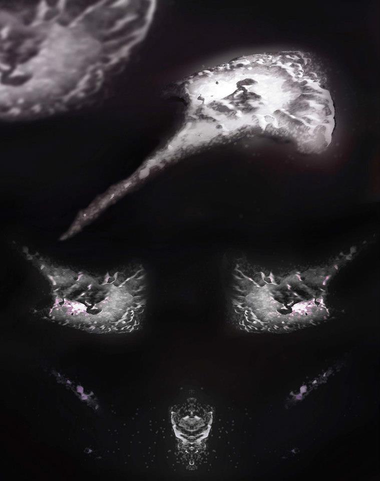
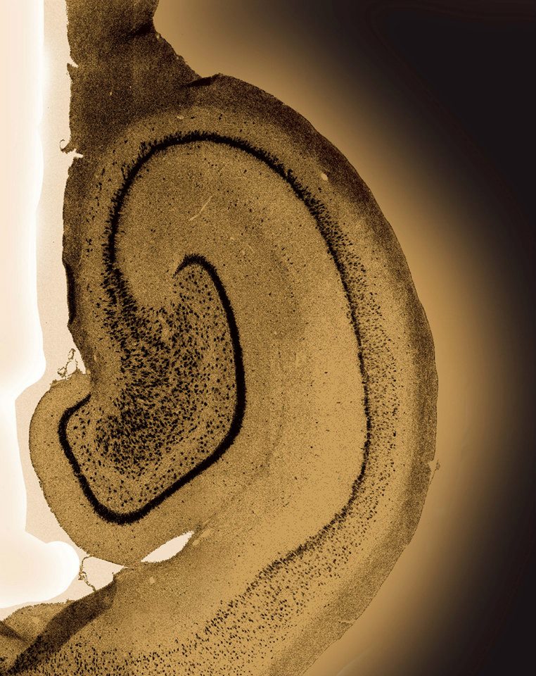
This image shows the brain of a piglet, which is used as a model to understand human brain development. The folded area shown is the hippocampus, an area important for learning and memory. Scientists are looking at how factors such as early-life nutrition or disease affect the development of the hippocampus, in an effort to discover how nutrition might protect the developing brain from harmful effects of inflammation.
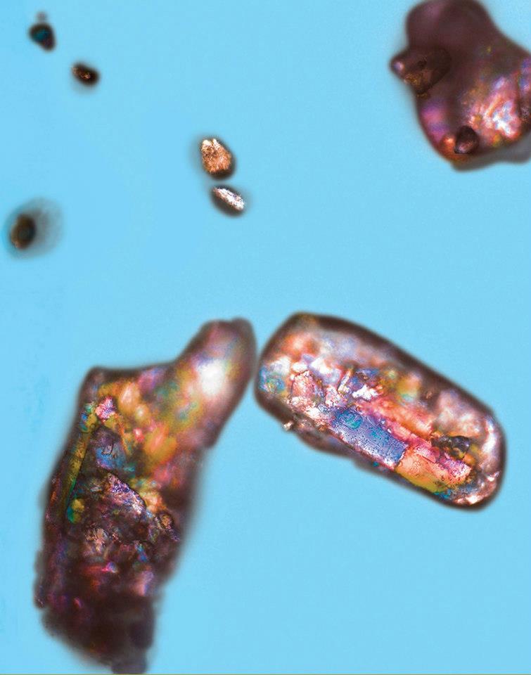
When crystals of glucose, a form of sugar used in many foods, absorb water, the way they refract light changes. Using polarized light microscopy, this change can be quantified, allowing scientists to study how and why glucose clumps or cakes during storage. Preventing the caking of glucose in a bag of sugar at home or silo of sugar in a factory could help ensure quality and prevent waste.
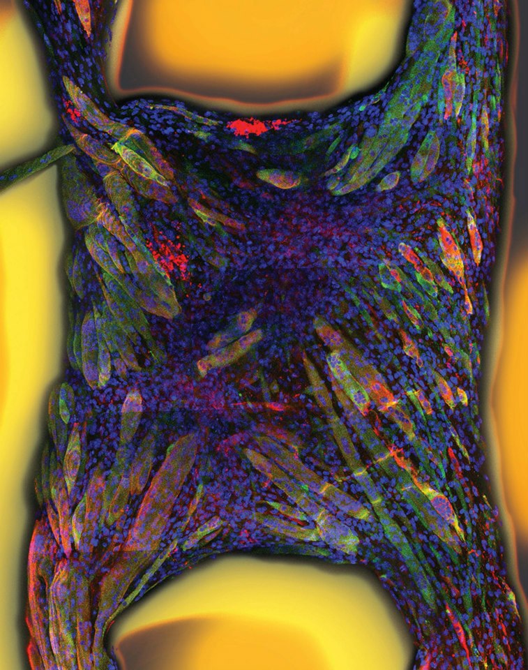
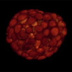
Here, the researchers have documented a way to coax stem cells to mature and form skeletal muscle in a matrix bed of proteins. Within two weeks, the resulting muscle fibers are able to contract.
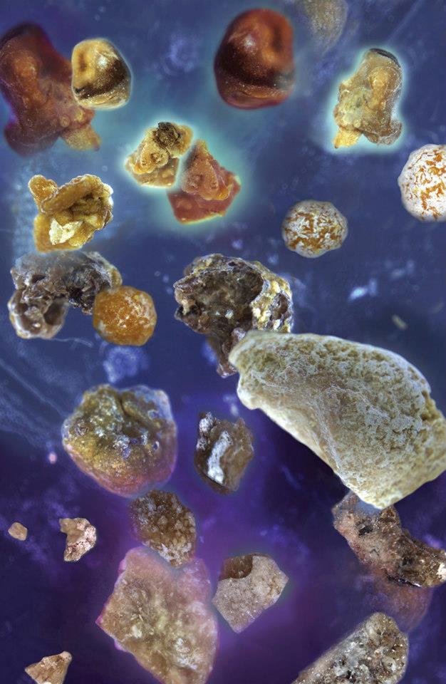
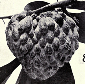
Human kidney stones exhibit a wide variety of shapes and forms, from rounded grape-like clusters to sharp and jagged glass-like shards. A collection of stones from different individuals demonstrates this great diversity of form.
Image provided by Yunzi Luo, Huimin Zhao Lab
Research funded by National Academies Keck Futures Initiative on Synthetic Biology
This image represents the energy released by the protons in a molecule extracted from bacteria, which is detected by sweeping the molecule with radio frequency in the presence of a magnetic field. The spectrum gives the researchers information about the connections between the atoms that make up the molecule, revealing its chemical structure.
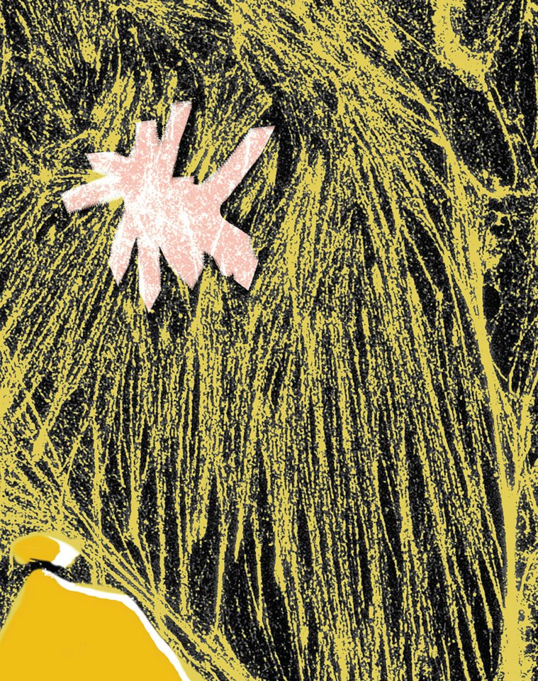
This image of the internal structure, or cytoskeleton, of a cell was produced through super-resolution microscopy. This new technique defies the physical limits of conventional microscopy and reveals twice the detail that can be obtained with older techniques. The higher level of detail helps researchers understand the structure and mechanics of much smaller objects.
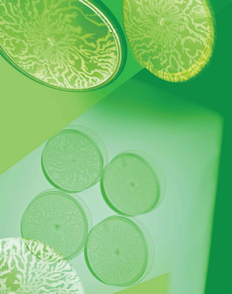
Some types of bacteria exhibit swarming motility: the ability to travel quickly as a group, due to coordinated movement that causes the group to flow across a surface. This movement causes the branching, fractal pattern seen here in round laboratory dishes. Researchers in the Enzyme Function Initiative work to discover the function of unknown proteins; in this case, a protein involved in swarming motility.
Bio-bot on the move
Scientist Collaborator
Vincent Chan
Rashid Bashir Lab
Instrument
Canon EOS 5D Mark II Digital SLR Camera
Funding Agency
Research funded by the National Science Foundation
Original Imaging
Special Thanks
Proton Minuet
Scientist Collaborator
Yunzi Luo
Huimin Zhao Lab
Instrument
Agilent 600 MHz NMR
Funding Agency
Research funded by National Academies Keck Futures Initiative on Synthetic Biology
Original Imaging
Special Thanks
Coral communities
Scientist Collaborator
Carly Hill Miller
Mayandi Sivaguru
Glenn Fried
Bruce Fouke
Bruce Fouke Lab
Instrument
Zeiss LSM 710 two photon microscope
Funding Agency
Research funded by the Office of Naval Research
Original Imaging
Special Thanks
World in a grain
Scientist Collaborator
Luke Mander
Surangi Punyasena Lab
Instrument
Zeiss Apotome, Zeiss LSM 710 NLO
Funding Agency
Research funded by the National Science Foundation
Original Imaging
Special Thanks
Rivers of life
Scientist Collaborator
Sinan Arslan
Taekjip Ha Lab
Instrument
Asylum Cypher High Resolution AFM
Funding Agency
Research funded by the National Institutes of Health, the National Science Foundation, and the Howard Hughes Medical Institute
Original Imaging
Special Thanks
Powerhouses of the cell
Scientist Collaborator
Vladimir Kolossov
Rex Gaskins Lab
Instrument
ELYRA superresolution microscope
Funding Agency
Research funded by National Institutes of Health
Original Imaging
Special Thanks
Nursery colors
Scientist Collaborator
Tim Abbott
Manabu Nakamura Lab
Instrument
Zeiss LSM 700 Confocal Microscope
Funding Agency
Research funded by Martek Biosciences Inc. and the United States Department of Agriculture
Original Imaging
Special Thanks
Zeiss Elyra
Scientist Collaborator
Tim Abbott
Manabu Nakamura Lab
Instrument
Zeiss Elyra SR-SIM Superresolution system
Funding Agency
Research funded by the Institute for Genomic Biology
Original Imaging
Special Thanks
Growing appetite
Scientist Collaborator
Juan Castro
James Drackley Lab
Instrument
NanoZoomer Slide Scanner
Funding Agency
Research funded by Milk Specialties Global
Original Imaging
Special Thanks
Invasion’s forefront
Scientist Collaborator
John Paul Eichorst
Robert Clegg
Glenn Fried
Yingxiao Wang
Robert Clegg Lab
Instrument
Multiphoton Confocal Microscope Zeiss 710 with Mai Tai eHP Ti: sapphire laser and Imaris
Funding Agency
Research funded by National Institutes of Health
Original Imaging
Special Thanks
Through a mathematical lens
Scientist Collaborator
Mayandi Sivaguru
Core Facilities
Instrument
Zeiss Elyra S1: SR-SIM System
Funding Agency
Research funded by the Institute for Genomic Biology
Original Imaging
Special Thanks
The finer point
Scientist Collaborator
James Zhu
Shiv Gopal Kapoor
Shiv Gopal Kapoor Lab and Chao-Trigger Machine Tool Systems Lab
Instrument
Zeiss LSM 700 Confocal Microscope
Funding Agency
Research funded by the Grayce Wicall Gauthier Chair in the UIUC MechSE department
Original Imaging
Special Thanks
Food for thought
Scientist Collaborator
Matthew Conrad
Rodney Johnson Lab
Instrument
Zeiss LSM 700 Confocal - four laser point scanning confocal with a single pinhole
Funding Agency
Research funded by the National Institutes of Health and the United States Department of Agriculture
Original Imaging
Special Thanks
Light of sweetness
Scientist Collaborator
Sarah K. Scholl
Shelly J. Schmidt Lab
Instrument
Fluorescence Microscope - Zeiss Axiovert 200M with the Apotome
Funding Agency
Research funded by various donor funds
Original Imaging
Special Thanks
Gaining strength
Scientist Collaborator
Vincent Chan
Rashid Bashir Lab
Laboratory of Integrated Biomedical Micro/Nanotechnology & Applications
Instrument
Multiphoton Confocal Microscope Zeiss 710 with Mai Tai eHP Ti: sapphire laser
Funding Agency
Research funded by the National Science Foundation
Original Imaging
Special Thanks
Cosmic kidney stones
Scientist Collaborator
Jim Bruce
Joe Weber
Sheila Egan
Yiran Dong
Bruce Fouke
Bruce Fouke Lab
Instrument
Microscope Zeiss Axiozoom V16 Scope
Funding Agency
Research funded by the National Science Foundation
Original Imaging
Special Thanks
Inner Light
Scientist Collaborator
Mayandi Sivaguru
Core Facilities
Instrument
Zeiss LSM 710 Two photon Microscope
Funding Agency
Research funded by Novus International Inc.
Original Imaging
Special Thanks
Cellular frontier
Scientist Collaborator
Mayandi Sivaguru
Core Facilities
Instrument
Zeiss Elyra Structured Illumination Superresolution System
Funding Agency
Research funded by the Institute for Genomic Biology
Original Imaging
Special Thanks
Go with the flow
Scientist Collaborator
Jose Solbiati
John Gerlt Lab
Enzyme Function Initiative
Instrument
Canon EOS 5D Mark II Digital SLR Camera
Funding Agency
Research funded by National Institutes of Health
Original Imaging
Special Thanks
Champaign businessman Doug Nelson, President of BodyWork Associates, first proposed the idea that became Art of Science, and his continued efforts to support the exhibit made its realization possible. The IGB is also grateful to James Barham of Barham Benefit Group and [co][lab] founder Matt Cho for hosting the annual exhibit.