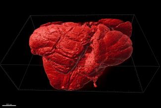
Half-Mouse Brain: This tile scan of a whole brain of a cuprizone-fed mouse was obtained with an Ultra Microscope (a 2nd generation lightsheet microscope by Miltenyi Biotec) after CLARITY tissue clearing and subsequent staining with anti-proteolipid protein (PLP). The image was processed with the Imaris 3D Software System: UltraMicroscope II - Miltenyi Biotec. Sample provided by Allison Yukiko Louie, Dr. Steelman's Laboratory, Department of Animal Sciences and imaged/proceed by Dr. Kingsley Boateng, Carl R. Woese Institute of Genomic Biology, UIUC.
