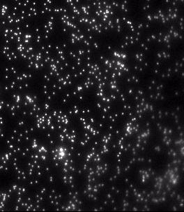
It has been shown that exposure to green (λ=550 nm, I=16 W/m2) fluorescent excitation light can induce force relaxation in living cells. Minimizing illumination time is critical to reducing light-induced force relaxation. We utilized a microscopic setup equipped with a mechanical shutter to evaluate the effect of short (<1s) exposure of living fibroblast cells to green light for one hour following the initial exposure. Cells were plated on polyacrylamide hydrogels embedded with dark red fluorescent nano particles which serve as fiducial markers. Motion of the nano particles was used to quantify change in cell force. Using a Zeiss Apotome Fluorescent Microscope with a Zeiss Axioobserver Z1 built-in reflected light shutter (<60 ms frequency), we exposed cells to green (λ=550 nm, I=16 W/m2) fluorescent excitation light for t=500 ms. Following this initial exposure, an image of the beads under each cell was captured every 5 minutes for one hour with dark red fluorescent excitation light (λ=650 nm, I=8.6 W/m2), which does not induce force changes. A widefield fluorescent metal halide lamp (X-Cite® Series 120Q) was used in conjunction with Semrock Brightline FP (green) and Cy5 (red) filters to illuminate the sample. The video shows the one-hour time lapse sped up 3000x (0.1s = 5 min in real time). The bead motion in the video illustrates that cells exposed to green light for only 500 ms do not exhibit widespread relaxation following exposure. The inward-outward-inward bead fluctuations show that localized cell forces continue to oscillate long after exposure to green fluorescent excitation light. Reducing exposure time to 500 ms may eradicate the widespread, light-induced force relaxation commonly observed in cells following initial illumination.
Watch the Video